Chapter 6: Basic radiation detectors
Inorganic scintillator (light quantum) 25: Inorganic scintillator + PMT (electron) 100: Inorganic scintillator + Si diode (electron–hole pair) 35 ...
Download Chapter 6: Basic radiation detectors

Information
Domain:
Source:
Link to this page:
Documents from same domain

Chapter 11: Computed Tomography - Human Health Campus
humanhealth.iaea.orgClinical Computed Tomography (CT) was introduced in 1971 -limited to axial imaging of the brain in neuroradiology It developed into a versatile 3D whole body imaging modality for a wide range of applications in for example • oncology, vascular radiology, cardiology, traumatology and interventional radiology. Computed tomography can be used for

Chapter 12:Physics of Ultrasound - Human Health Campus
humanhealth.iaea.orgDiagnostic Radiology Physics: a Handbook for Teachers and Students –chapter 12,11 12.2. ULTRASONIC PLANE WAVES 12.2.3. Reflection and Transmission An ultrasound image displays the magnitude (absolute value of amplitude) of ultrasound echoes , so a physical understanding of acoustic wave reflection is valuable for interpreting the images

Chapter 5:X-Ray Production - International Atomic Energy ...
humanhealth.iaea.orgInternational Atomic Energy Agency Slide set of 121 slides based on the chapter authored by R. Nowotny of the IAEA publication (ISBN 978-92-0-131010-1): Diagnostic Radiology Physics: A Handbook for Teachers and Students Objective: To familiarize the student with the principles of X ray production
Chapter 8:Fluoroscopic Imaging Systems
humanhealth.iaea.org0.9-0.95 for fixed-pixel devices (e.g. CCD cameras) and liquid crystal display (LCD) monitors In the Horizontal direction, resolution is limited by the bandwidth of the video system In most systems the bandwidth is adjusted to give Equal Resolutionin both the vertical and horizontal directions 8.2 FLUOROSCOPIC EQUIPMENT
Finding Early Invasive Breast Cancers: APracticalApproach
humanhealth.iaea.orgnvasive breast cancer typically man-ifests as a mass at mammography. Before developing into a mass, a can-cer may manifest as a focal asymmetry. Early detection of these masses and asymmetries that represent invasive carcinoma is important in reducing breast cancer mortality. Women with invasive cancers of 1 cm or smaller have

Chapter 13: Image Reconstruction
humanhealth.iaea.org13.2.1 Two dimensional tomography Eq. 13.1 describes the acquisition process in 2-D PET and in SPECT with parallel hole collimation, if attenuation can be ignored. Assuming that Λ(x, y) represents the tracer distribution at transaxial slice . Z. through the patient, then Y(s,ϕ) represents the corresponding sinogram, and contains the . z
Related documents
Time-of-Flight Mass Spectrometry - Agilent
www.agilent.comScintillator Figure 2. TOF detector with potentials shown for positive ion operation. The reason for this conversion of an electrical signal to an optical signal and back to an electrical signal is to electrically isolate the flight tube and the front of the detec-tor, which are at roughly –6,500 volts, from the PMT, whose signal output is at
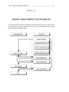
IMAGE PROCESSING TECHNIQUES
users.ncsa.illinois.edub) scintillator-photomultiplier combination which is used for recording secondary electrons. The secondary electrons are detected by a scintillator-photomultiplier combination which is known as the Everhart-Thornley detector. Its construction is shown in Fig. 1.1.2b which is taken form [1.1.2]. The secondary electrons are collected by a grid.
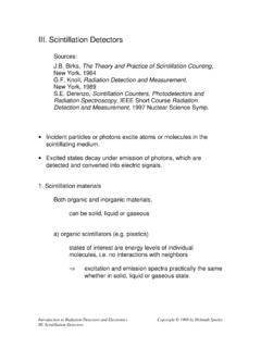
III. Scintillation Detectors
www-physics.lbl.govFor an ideal scintillator and low ionization density Luminescence ∝ Energy dissipated in scintillator or, in differential form The specific density of ionized and excited molecules along the particle track is Assume that a portion of the primary excitation is lost at high ionization density (ionization quenching) and introduce a quenching ...

X-ray detectors - Hamamatsu
www.hamamatsu.comScintillator detectors are widely used in these applications. These detectors use scintillators to convert X-rays into light and detect this light to detect X-rays indirectly. Especially in the medical field, the digital X-ray method, which uses X-ray detectors with large photosensitive area, is becoming mainstream, replacing the
Gamma Ray Spectroscopy - Department of Physics
www.phys.ufl.eduwith the scintillator; many do not interact at all and simply pass right through. More-over, there can be one or more interactions of a gamma and there may be multiple gam-mas emitted from a single nuclear decay. Usu-ally, all energy depositions in the scintillator from a single decay are effectively simultane-
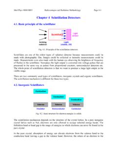
Chapter 4 Scintillation Detectors
www.science.mcmaster.cascintillator in use. The average quantum efficiency over the emission spectrum of a typical scintillator is about 15 to 20 percent while the peak quantum efficiency is 25 ~ 30 %. The standard for quotation is the number of photoelectrons per keV energy loss …

The Decay of Muons - UCL
www.ucl.ac.ukThe scintillator and photomultiplier tube (PMT) were both installed inside a black anodized aluminium cylinder with measurements of ( ) diameter and ( ) height, as shown in Fig. (3). [9] Polyvinyltoluene served as the base material for the scintillator and was shaped as a cylinder of ( …