
Clinical Practice Guidelines
volving collateral ligament of knee, Sprain and strain involving cruciate ligament of knee, and Injury to multiple structures of knee, and the associated International Classification of Functioning, Dis-ability and Health (ICF) impairment-based categories of knee insta-bility (b7150 Stability of a single joint) and movement coordination
Download Clinical Practice Guidelines

Information
Domain:
Source:
Link to this page:
Documents from same domain

Neck Pain: Clinical Practice Guidelines Revision 2017
www.orthopt.orgNeck Pain: Clinical Practice Guidelines Revision 2017 tion to neck pain. Overall, classification is critical for match - ing the intervention strategy that is most likely to provide
Heel Pain—Plantar Fasciitis: Clinical Practice Guidelines ...
www.orthopt.orgHeel Pain—Plantar Fasciitis: Clinical Practice Guidelines Revision 2014 Key Clinical Findings of Heel Pain/Plantar Fasciitis • Plantar medial heel pain: most noticeable with initial steps after a period of inactivity but also worse following prolonged weight bearing (B)

CURRENT CONCEPTS IN FUNCTIONAL CAPACITY …
www.orthopt.orgCurrent Concepts in Functional Capacity Evaluation: A Best Practices Guideline Adopted April 30, 2018 Page 2 of 43

Neck Pain: Revision 2017
www.orthopt.orgthoracic segmental mobility tests; (2) neck pain with headache, in-cluding cervical active ROM, the cervical flexion-rotation test, and upper cervical segmental mobility testing; (3) neck pain with radiat-ing pain, including neurodynamic testing, Spurling’s test, the distrac-tion test, and the Valsalva test; and (4) neck pain with movement
Adhesive Capsulitis: Clinical Practice Guidelines
www.orthopt.orgdeficits/adhesive capsulitis R ule in if: • Patient’s age is between 40 and 65 years • Patient reports a gradual onset and progressive worsening of pain and sti‘ness • Pain and sti‘ness limit sleeping, grooming, dressing, and reaching activities • Glenohumeral passive range of motion (ROM) is limited in multiple directions,
Clinical Instability of the Lumbar Spine: Diagnosis and ...
www.orthopt.orgspinal manipulation, provides preliminary evidence and is a stepping stone toward ran-domized clinical trials. The CPR is designed to assist clinicians in making better decisions regarding match-ing patients with the most appropriate inter-vention. The steps in creating a clinical pre-diction rule are to: ( ) identify factors that

Ankle Stability and Movement Coordination Impairments ...
www.orthopt.orgANKLE SPRAIN B Clinicians should use special tests, including the reverse anterolateral drawer test and anterolateral talar palpation in addition to the traditional anterior drawer test, and a thorough history and physical examination to aid in the diagnosis of a LAS. DIAGNOSIS/CLASSIFICATION – CHRONIC ANKLE INSTABILITY
Related documents

Normal MR Imaging Anatomy of the Knee - SmartView
www.smartview.coof the MCL, and is elongated in a vertical fashion. The semimembranosus-tibial collateral ligament bursa, an inverted U-shaped structure, does not communicate with the joint, and is positioned between the semimembranosus tendon and tibial collateral ligament at the level of the medial tibial plateau.11 Laterally, the iliotibial band and fibular

Basic Knee Physical Exam Checklist 4.21
meded.ucsd.eduValgus stress test for MCL: Push on lateral aspect of knee while pulling ankle away from midline. Perform in 0 degrees (full extension) and 30 degrees flexion to try to isolate the medial collateral ligament Varus stress test for LCL: push on medial aspect of knee while pulling ankle towards midline. Perform in 0

Musculoskeletal Examination: General Principles and ...
meded.ucsd.eduSpecifics of Testing –Medial Collateral Ligament (MCL) •Flex knee ~ 300 •Left hand on lateral aspect knee. •Right hand on ankle or calf. •Push inward w/left hand (Valgus force). •If MCL torn, joint "opens up" along medial aspect. •May also elicit pain w/direct palpation over ligament Compare w/non-affected side –“normal”

MEDIAL COLLATERAL LIGAMENT (MCL) …
universityorthopedics.comMEDIAL COLLATERAL LIGAMENT (MCL) RECONSTRUCTION/ REPAIR POST-OP REHABILITATION PROTOCOL The following is a protocol for postoperative patients following ACL reconstruction. The primary goal of this protocol is to protect the reconstruction while steadily progressing towards and ultimately achieving pre-injury level of activity.
Rehabilitation Protocol for Medial Collateral Ligament Injury
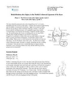
www.massgeneral.orgRehabilitation after Injury to the Medial Collateral Ligament of the Knee Phase 2: Six weeks after injury onward (grade 2 and 3) Three weeks after injury onward (grade 1) This handout is to help you rebuild the strength of the knee muscles after injury to the MCL of the knee. It is intended as a guideline to help you organize a structured ...

Rehabilitation Guidelines for Knee Multi-Ligament Repair ...
www.uwhealth.orgthe medial collateral ligament (MCL) (Figure 1a), lateral collateral ligament (LCL) (Figure 1b), anterior cruciate ligament (ACL) (Figure 2a) and posterior cruciate ligament (PCL) (Figure 2b). The MCL connects the femur and tibia medially (on the inside) and resists valgus (knee buckling in) knee motion. A common mechanism of injury

CODING ARTHROSCOPIC KNEE PROCEDURES
www.velocityhc.com• The Medial Collateral Ligament (MCL). The MCL is located on the inside (medial side) of the knee, connecting the inside, bottom edge of the femur with the inside, top edge of the tibia. The MCL helps to stabilize the knee by limiting inward (valgus) force across the knee. The MCL works with the LCL to prevent unwanted side-to-side motion.