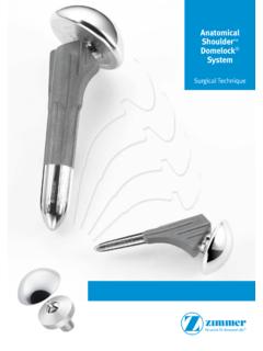
Transcription of ZIMMER BIGLIANI/ FLATOW THE COMPLETE …
1 ZIMMER . bigliani /. FLATOW . THE COMPLETE . shoulder . SOLUTION. Total shoulder Arthroplasty Surgical Technique BIGLIANI/ FLATOW CONTENTS. THE COMPLETE SURGICAL TECHNIQUE FOR BIGLIANI/ FLATOW . shoulder SOLUTION shoulder SYSTEM. SURGICAL TECHNIQUE Patient Positioning .. 2. Incision and Exposure .. 2. Humeral Preparation .. 3. Instruments and surgical technique developed in conjunction with: Glenoid Preparation .. 9. Humeral Head Selection .. 15. Louis bigliani , Expanded Glenoid Options .. 16. Chairman, Department of Orthopaedics Implantation .. 18. Director, The shoulder Service Humeral Head Removal.
2 25. New York Presbyterian Hospital Closure .. 25. Columbia-Presbyterian Center Postoperative Management .. 25. New York, New York Evan FLATOW , Chief of shoulder Surgery Mount Sinai Medical Center New York, New York 1. PATIENT POSITIONING. Patient positioning is especially important in total shoulder surgery (Fig. 1). Place the patient in a semi- beach-chair position. Use a head rest that allows for the superior part of the table to be removed. Place two towels under the spine and the medial border of the scapula to raise the affected side. Raise the head of the table approximately 25-30 degrees to reduce venous pressure.
3 Attach a short arm board to the table, or use another arm support method that will allow the arm to be raised or lowered as necessary throughout the procedure. Fig. 3. expose the deltoid and pectoralis major muscles. Retract the skin by placing Gelpi retractors about one- third of the way down, and one-third of the way up. Develop the deltopectoral interval, retracting the pectoralis major medially and the deltoid laterally. Fig. 1 Identify and dissect the interval between the pectoralis major muscle and the cephalic vein. Retract the ceph- INCISION AND EXPOSURE alic vein either medially or, preferably, laterally as this First, mark the coracoid process.
4 Then mark the line will minimize bleeding from the deltoid muscle or of the incision, beginning at the clavicle just lateral to cephalic vein. Release the upper 1cm-2cm of the the coracoid process. Extend the line along the del- insertion of the pectoralis major tendon, being careful topectoral groove to the area of the mid-humerus (Fig. to avoid the long head of the biceps tendon. In very 2). Then make the incision following the line. Under- tight shoulders, the pectoralis may need to be com- mine the skin flaps to improve exposure. Dissect pletely released.
5 Tag the pectoralis major muscle with subcutaneous tissue from the deltoid fascia, and a suture so it can be easily identified for later reattach- ment. Reposition the distal medial retractor under the pectoralis major muscle. Release any adhesions between the deltoid and strap muscles (coracobrachialis and short head of biceps). and develop a plane between the strap muscles and the humerus. Reposition the proximal medial retractor under the strap muscles (Fig. 3). Sweep the bursa off the humeral head and the greater tuberosity. Then expose the superior portion of the subacromial space by resecting the leading edge of the coracoacromial ligament.
6 Identify the superior and inferior margins of the subscapularis tendon. Divide the tendon just medial to the bicipital groove and remove it from the lesser tuberosity. Being careful inferiorly to avoid the axillary nerve, retract the sub- scapularis medially, exposing the articular surface of Fig. 2. the humeral head. 2. It is very important to maintain as much length of the REAM HUMERAL CANAL. subscapularis muscle as possible. Remove the cap- Dislocate the humeral head by externally rotating and sule and subscapularis tendon as a unit laterally from extending the humerus.
7 If necessary, place a metal the humerus. Do this as close as possible to the hu- finger inferiorly between the humerus and the glenoid. meral neck to avoid injury to the axillary nerve. Exter- Removal of capsule from the inferior aspect of the nal rotation of the humerus is helpful. In cases where humeral neck may be needed to achieve dislocation. there is a large inferior osteophyte on the humeral Before reaming the canal, it is important to remove all head, it is especially important to dissect the capsule anterior or inferior osteophytes so that the true ana- off the neck of the humerus laterally as the axillary tomical neck (junction of the articular cartilage and nerve is on the medial and inferior aspect of the os- cortical bone) can be determined.
8 Teophyte. Medially, at the glenoid rim, the capsule and labrum can be separated from the subscapularis Attach a Short Intramedullary Reamer with a trocar tendon. This will facilitate lateral mobilization. point to the Ratchet T-handle. There are three posi- tions marked on the collar of the T-handle: FORWARD, LOCKED, and REVERSE. To ream the starter hole, use HUMERAL PREPARATION the FORWARD position. Short Intramedullary Reamers The goal in replacing the humeral head is to place a are available in diameters of 6mm, 7mm, and 8mm, prosthetic articular surface precisely on the proximal and the appropriate size should be chosen for the humerus as it would have been before the destructive patient's humeral canal.
9 Place the trocar tip of the arthritic process began. The relationship among bony reamer just posterior to the bicipital groove (Fig. 4). anatomy, rotator cuff insertions, and soft tissue ten- and ream a starter hole. A mallet may be used to start sion must all be considered. the hole in hard bone. Technique Tip: To facilitate access to the humeral canal, the shoulder should be off the table. To accomplish this, push the elbow back and externally rotate the arm. Short Reamer Ratcheting T-Handle Fig. 4. 3. Attach the T-handle to the longer, blunt-tipped In- Note: If using a 60mm length stem, use only the tramedullary Tapered Reamer of the same diameter Short Intramedullary Reamers.
10 Use the and begin manually reaming the humeral canal. Use Intramedullary Tapered Reamers for 110mm progressively larger reamers in 1mm increments until monoblock stems. Ream until the flutes are resistance is felt from cortical contact in the canal buried in the bone. (Fig. 5). Continue reaming to the appropriate depth as indicated on the reamer shaft. The depth corresponds to the implant length chosen. Do not remove cortical bone. These reamers have blunt tips to help guide them down the canal and prevent obtrusion into cortical bone. Remove the T-handle, but leave the last reamer in the canal to interface with the Humeral Head Cutting Guide (Fig.)