Transcription of The Digestive System PDF - Hart County, Georgia
1 1 The Digestive SystemThe Digestive System Jim SwanThese slides are from class presentations, reformatted for static viewing. The content contained in these pages is also in the Class Notes pages in a narrative format. Best screen resolution for viewing is 1024 x 768. To change resolution click on start, then control panel, then display, then you are viewing this in Adobe Reader version 7 and are connected to the internet you will also be able to access the enriched links to notes and comments, as well as web pages including animations and videos. You will also be able to make your own notes and comments on the pages. Download the free reader from Discussed in the Digestive System :Ingestion food intakeMastication chewing (provides mechanical or physical digestion)Deglutition swallowingPropulsion (motility) movement through Digestive tract via muscular (transverse) muscle segmentationlongitudinal muscle peristalsisSecretion release of mucus, enzymes and other substances along with associated with the Digestive System : ingestion- intake of food, mastication- chewing, a component of physical digestion, deglutition swallowing, propulsion- movement of materials along the alimentary canal.
2 Occurs mostly in the alimentary canal as muscular movements producing segmentationand peristalsis, secretion- the release of substances from cells in the Digestive tract, mucus, enzymes, hormones, etc. 3 Digestive Processes (contd.)Absorption the transport of Digestive endproducts into the blood or the shedding of mucosal lining elimination of waste containing bacteria, exfoliated cells, and undigested digestion breaks down food mass exposing food to enzymeschemical digestion enzymatic hydrolysis which breaks complex molecules into their the transport of Digestive endproducts into the blood or the lymph; defecation- the removal of waste from the GI tract including undigested materials, exfoliated cells and bacteria; exfoliation-the constant shedding of the mucosal lining cells and their replacement by mitosis; digestion- consists of physical (mechanical) digestion, and chemical digestion; Physical digestion is the reduction in bulk and increase in surface area of ingested food.
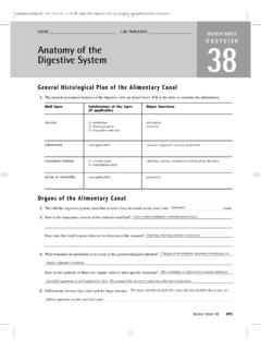
3 ; Chemical digestion is enzymatic hydrolysisin which large complex molecules are broken down to their subunits. Physical digestion makes chemical digestion possible. 4 The Digestive TractThe alimentary canalis a continuous tube stretching from the mouth to the , sublingual, and submaxillary salivary glands. There are also buccal cells in cheek which secrete Gallbladder StomachEsophagusPancreas Colon (largintestine)Small intestineRectumAnusThe Digestive tract is composed mostly of the alimentary canal (see next frame), together with accessory glands and organs. The alimentary canalis the continuous tube stretching from the mouth to the anus. Components of this tube, the various organs of the System , are specialized to perform particular functions. The stomach and intestines are commonly referred to as the GI (gastrointestinal) tract, but this designation is also often used to include the entire alimentary Alimentary Canal CSFibro-serous covering MMyenteric nerve plexusSubmucosal nerve plexusMuscularis (externa): longitudinal transverse (circular)Submucosa: contains glands and blood vesselsMuscularis mucosae: Delineates the mucosaMucosa: includes the epithelial lining and lamina propria Covering is fibrous in esophagus, serous in most of GI tract.
4 Serous membranes also form mesenteries and the greater and lesser is fibrous in esophagus, serous in most of GI tract. Serous membranes also form mesenteries and the greater and lesser alimentary canalis composed of four layers, each layer typically composed of certain tissues. But these layers can vary somewhat within the canal. mucosa- this is the lining tissue, mostly made of simple columnar epithelium (the mucosa of the esophagus is non-keratinized stratified squamous epithelium). Goblet cells within this layer secrete mucus for lubrication and protection and other cells may secrete enzymes, hormones etc. The lining through much of the alimentary canal exfoliates on a 3 to 5 day cycle. The gastrointestinal mucosa is also responsible for absorption of Digestive endproducts. Beneath the epithelial surface is a connective-like component called the lamina propria. This layer contains blood and lymph capillaries for absorption.
5 The boundary of the mucosa is the muscularis mucosae, the "muscle of the mucosa" which contracts to increase exposure of the mucosal lining to contents of the alimentary canal. The submucosa-this layer lies beneath the mucosa and is basically areolar connective tissue containing major blood vessels, nerves, and lymph nodes serving the alimentary canal. The submucosal nerve plexus controls the function of mucosal cells and Digestive functions. The muscularis(or muscularis externae) - this is mostly smooth muscle (the esophagus has partly skeletalmuscle) in two or three layers. In most of the GI tract two layers exist, the longitudinal smooth musclelayer and the circularor transverse smooth muscle layer. The circular layer squeezes to produce segments in the intestines, while the longitudinal layer causes the repeated shortening and lengthening called peristalsis . Segmentation contractions are mostly mixing actions, but work together with peristalsis in propulsion.
6 The serosa or fibroserous layer- this is the covering, a serous membrane in the portions of the alimentary canal in the peritoneal cavity and a fibrous covering in portions not in the peritoneal cavity or considered retroperitoneal. 6 The alimentary canal consists of four layers. Listed from outside to inside: 1) the fibroserousouter covering; serous in most of GI tract, fibrous in esophagus. 2) the muscularis(externa), smooth muscle (except in upper esophagus) in two or three layers; 3) the submucosa, containing glands, nerves, and blood vessels; 4) the mucosa, the epithelial secretory and absorptive lining, bounded by the muscularis mucosae (mm).1234mmWall of the Alimentary CanalOrgans and Regions of the Alimentary Canal: (See previous frame)Note that in this view the lining is shown with villi. This arrangement is seen in the small PeritoneumPeritoneal organs are covered by serosa, the visceral peritoneum. The peritoneal cavity is lined by the parietal organs have fibrous covering on part of omentumMesenteries Small intestineUrinary bladderRectum Figure dDuodenum Pancreas Stomach Transverse colonThe peritoneal cavity (blue area in above slide) houses most of the Digestive organs.
7 It is lined with a serous membrane, the parietal peritoneum, which is continuous with the visceral peritoneumthat covers these organs. Within the abdomino-pelvicregion there are also organs not in the peritoneal cavity or retroperitoneal, which are covered with fibrosarather than omentumLiver Stomach Lesser omentumTransverse mesocolonTransverse colonJejunum Ileum Sigmoid colonThe mesenteriesare double layers of serous membrane, composed of peritoneal membranes which have folded against each other. Thesemesenteries connect and hold gastrointestinal organs in place and attach blood vessels and nerves. They also, with their fatty coverings, protect and insulate the organs. The greater omentum, for instance, hangs in front of the intestines acting as an insulator and shock MesenteriesSeen here is a loop of bowel attached via the mesentery. Note the extent of the veins and arteries. There is an extensive anastomosing arterial blood supply to the bowel, making it more difficult to infarct.
8 Also, the extensive venous drainage is incorporated into the portal venous System heading to the Chart: The MouthAREAPROCESSESSECRETIONSCON TRO LSHISTO LOGYM outhmechanical digestionchemical digestion: starches->shorter chainssaliva:salivary amylase(ptyalin)cephalicphysicalcontactn on-keratinizedstratifiedsquamous;salivar y glandsSaliva also contains water, electrolytes, mucus and serous fluid, also contains water, electrolytes, mucus and serous fluid, and chemo-receptors respond to presence of substances in the and chemo-receptors respond to presence of substances in the mouth:The mucosa of the mouth is composed of mostly non-keratinized stratified squamous epithelium. This mucosa continues through the esophagus. Three salivary glands on each side, plus buccal glands in the mucosa, provide the fluid known as saliva. Saliva contains water, salts, mucin, serous fluid, lysozyme, IgA, growth factors, and amylase.
9 11 The Oral CavityFigure glandParotid duct Masseter Submandibular glandSubmandibular ductSublingual glandSublingual ductsBuccal glands, groups of salivary glandular cells, also line the glands, groups of salivary glandular cells, also line the three glands (parotid, submandibular, and sublingual) produce varying amounts of salivary components. The pH of this fluid is from to , supporting the action of salivary amylase to begin the breakdown of polysaccharides to shorter chains. The action does not normally progress very far due to the shortexposure to active enzyme. 12 DeglutitionThe muscle at the upper end of the esophagus functions like a sphincter, but it is not a structural muscle at the upper end of the esophagus functions like a sphincter, but it is not a structural esophageal muscle contractedbolusUvula lifts upUpper esophageal muscle relaxes, bolus is pushed into begins as a voluntary act, placing the bolus of food on the tongue and contracting tongue and pharyngeal begins as a voluntary act, placing the bolus of food on the tongue and contracting tongue and pharyngeal becomes involuntary when esophageal muscles contract initiating becomes involuntary when esophageal muscles contract initiating is a form of mechanical digestion which reduces the bulk of the food and, especially, exposes it to the enzyme.
10 The bolus of food is swallowed in a process called deglutitionwhich begins as voluntary and becomes involuntary. At first the bolus is lodged on the tongue and pushed voluntarily into the pharynx. Then pharyngeal muscles contract pushing the bolus into the esophagus where peristalsis begins. Peristaltic waves move food down the esophagus into the Along EsophagusThere is no structural gastroesophageal (or cardiac) sphincter! The esophageal muscles at the lower end of the esophagus remain tonically contracted until peristalsis brings the bolus of food. These muscles produce a functionalbut not a is no structural gastroesophageal (or cardiac) sphincter! The esophageal muscles at the lower end of the esophagus remain tonically contracted until peristalsis brings the bolus of food. These muscles produce a functionalbut not a dCircular muscles contract pushing bolus down Longitudinal muscles shorten passageway ahead of bolusPeristalsis begins in the esophagus and moves the bolus into the stomach.