Transcription of Staged Arthroscopic Rotator Cuff Repair with a Bridging ...
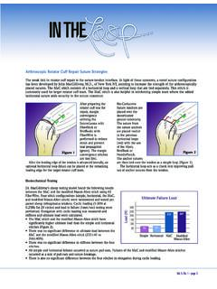
1 Staged Arthroscopic Rotator cuff Repair witha Bridging Acellular Human Dermal Graftin the Treatment of Infection FollowingOpen Rotator cuff RepairACaseReportVivek Agrawal, MDInvestigation performed at The Shoulder Center, Carmel, IndianaAlthough infection after Rotator cuff Repair occurs in-frequently, it can be a catastrophic complication. To thebest of my knowledge, there are only a small numberof reports in the literature regarding management of theseinfections, and none of these report the outcome of a stagedarthroscopic approach1-6. Once the infection has resolved, re-construction of the residual Rotator cuff defect can present asubstantial challenge for the treating orthopaedic purpose of this report is to present the clinical,functional, and magnetic resonance imaging (MRI) results ofan all- Arthroscopic approach for the management of a massiverotator cuff tear defect resulting from an infection after aninitial open Rotator cuff Repair .
2 The patient was informed thatdata concerning his case would be submitted for publication,and he provided ReportAthirty-two-year-old right-hand dominant man withmedical comorbidities including diabetes, hypertension,and obesity (body-mass index [BMI] > 40) was referred to ourshoulder clinic in October 2009 for a persistent deep leftshoulder infection after an open Rotator cuff Repair . The patienthad initially been treated with an open Rotator cuff Repair witha mesh reinforcement graft in August 2009. The patient hadexperienced persistent pain and had developed a drainingwound, which had been managed with serial debridement,wound care, and intravenous antibiotics for a period of threeweeks, followed by oral antibiotics (Augmentin [amoxicillinand clavulanate] and then Levaquin [levofloxacin]) and dressingchanges.
3 Previous culture specimens had been positive forPropionibacterium acnes. Despite the aggressive surgical care andserial debridement, the patient had a persistent arthrocutaneousfistula along with dehiscence of the deltoid muscle. He then wasreferred to our tertiary care shoulder clinic for definitiveFig. 1 MRI (coronal view) of the shoulder, consistent with osteomyelitis of thegreater tuberosity, diffuse infectious synovitis, deltoid dehiscence, ar-throcutaneous fistula, and a massive Rotator cuff tear with substantialretraction and fibrosis to the undersurface of the :The author received payments or services, either directly or indirectly ( , via his institution), from a third party in support of an aspect of thiswork. In addition, the author, or his institution, has had a financial relationship, in the thirty-six months prior to submission of this work, with an entity inthe biomedical arena that could be perceived to influence or have the potential to influence what is written in this work.
4 The author has not had any otherrelationships, or has engaged in any other activities, that could be perceived to influence or have the potential to influence what is written in this work. ThecompleteDisclosures of Potential Conflicts of Interestsubmitted by the authors are always provided with the online version of the 2012 BYTHEJOURNAL OFBONE ANDJOINTSURGERY,INCORPORATEDJBJSCaseConn ect2012;2:e30 In addition to a thorough history and physical ex-amination, we also obtained an infectious disease consultationand MRI of the left shoulder with intravenous (IV) gadoliniumenhancement. The MRI was consistent with osteomyelitis of thegreater tuberosity, diffuse infectious synovitis, deltoid dehiscence,arthrocutaneous fistula, and a massive Rotator cuff tear involvingFig.
5 2 Fig. (coronalview)oftheshoulderfollowinginiti aldefinitivedebridement, (sagittalview)oftheshouldershowsgrade-1f attyinfiltrationofthesupraspinatusmuscle witha positive tangent sign consistent with substantial atrophy, and grade-2 fatty infiltration of the infraspinatus 4-AFig. 4-BFig. 4-AMRI (coronal view) of the shoulder at one-year postsurgery shows the supraspinatus tendon at the level of the humeral head with the Bridging graft(arrows).Fig. 4-BMRI (sagittal view) of the shoulder at one-year postsurgery shows the intact Bridging graft (arrows).2 JBJS CASECONNECTORVOLUME2dNUMBER3dJULY11, 2012 STAGEDARTHROSCOPICROTATORCUFFREPAIR WITHABRIDGINGACELLULARHUMANDERMALGRAFTth e supraspinatus and infraspinatus tendons with substantialretraction as well as atrophy with fibrosis of the Rotator cuff tothe undersurface of the acromion (Fig.)
6 1). After the infectiousdisease consultation, we recommended discontinuation of allantibiotics for at least two weeks prior to the initial radicaldebridement portion of our recommended Staged approachin order to have the best chance of isolating an organism in theintraoperative culture specimens. We performed an extensivearthroscopic debridement, including global capsulotomy, bicepstenotomy (frayed), coring of prior suture anchor tracks withremoval of retained foreign body and placement of vancomycin-impregnated calcium paste (PRO-DENSE; Wright Medical),excision of the fistula, primary Repair of the deltoid dehiscencewith absorbable monofilament suture, and closure of the skinwound with monofilament nylon suture. All culture speci-mens were negative, and the patient was empirically startedon daptomycin for a six-week postoperative duration.
7 Hehealed uneventfully, and a repeat MRI of the left shoulderwith IV gadolinium enhancement demonstrated excellenthealing of the deltoid dehiscence and resolution of the oste-omyelitis (Fig. 2). At that time, he had a massive Rotator cufftear with grade-1 fatty infiltration of the supraspinatus musclewith a positive tangent sign for substantial atrophy, and grade-2 fatty infiltration of the infraspinatus muscle with substantialretraction and fibrosis (Fig. 3). Two months after the radicaldebridement, we proceeded with revision Arthroscopic repairof the massive Rotator cuff defect. Because the patient had adeficiency of a substantial portion of the supraspinatus ten-don, we elected to utilize an acellular dermal graft (AllopatchHD; Musculoskeletal Transplant Foundation [MTF]) to serveboth reinforcement and Bridging functions.
8 The postopera-tive course was uneventful. The patient maintained supinepassive and active motion for twelve postoperative weeks andthen gradually progressed to full upright active shouldermotion. At the one-year follow-up, MRIs showed an intactbridging graft (Figs. 4-A and 4-B) with maintenance of grade-1 fatty infiltration of the supraspinatus muscle and increasedgrade-3 fatty infiltration of the infraspinatus muscle (Fig. 5).No evidence of recurrent or indolent infection was noted. Thepatient was extremely pleased with the outcome and had fullactive shoulder motion (Video 1; see Appendix). The Constant-Murley Shoulder Outcome Score improved from 25 (pretreat-ment) to 78 at the one-year follow-up, with the strength subsetscore improving from 0 to kg.
9 The normalized Flexilevel Scaleof Shoulder Function score improved from 50 (pretreatment) to70 at the one-year after Rotator cuff Repair is an infrequently reportedbut potentially devastating complication. Reported rates ofinfection following Rotator cuff Repair range from open repairs and mini-open repairs. Thereare other potential complications stemming from infectionfollowing Rotator cuff Repair , including acute adhesive capsu-litis, chondrolysis, osteomyelitis, secondary osteoarthritis, andfailure of the Rotator cuff repair5,8. Traditionally, the key tosuccess is early detection, radical debridement, and long-termantibiotic therapy. Many authors report a mean of two-and-a-half serial procedures in concert with antibiotics to treat thisproblem2, after open Rotator cuff Repair in the studies inthe literature presents as two distinct populations: one acute orsubacute, presenting within ninety days of the index procedure,and the other a chronic infection typically referred and/ortreated at an interval greater than ninety days.
10 Delay in diag-nosis and treatment can result in poorer et al. outlined several factors associated with anincreased risk of infection after open Rotator cuff repair5. Chenet al. noted an increased rate (10%) of infection after openrotator cuff Repair in thirty diabetic patients9. Our patient alsohad diabetes as an identifiable risk factor. As noted by Mirzayanet al. and reflected in our report, the number of patients in ourexperience is too small to draw any meaningful conclusions;however, a thorough evaluation preoperatively can help identifypatients who are at risk for deep infection following shouldersurgery. Because there appears to be a lower rate of infectionassociated with Arthroscopic surgery than with an open ap-proach, these patients may benefit when it is possible to use anarthroscopic most common pathogens cited in Rotator cuff in-fections includeStaphylococcus aureus,Staphylococcus epi-dermidis,Propionibacterium acnes, and coagulase-negativestaphylococci.