Color Flow Ultrasound In Detection
Found 8 free book(s)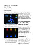
Doppler Color Flow Imaging #4 - Cardioland
cardioland.orgDoppler Color Flow Imaging #4 Joseph A. Kisslo, MD David B. Adams, RDCS INTRODUCTION Doppler color flow imaging is a method for noninvasively imaging blood flow through the heart
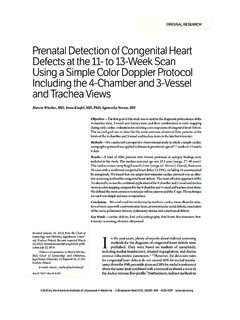
Prenatal Detection of Congenital Heart Defects at the 11 ...
www.dobreusg.plPrenatal Detection of Congenital Heart Defects at the 11- to 13-Week Scan Using a Simple Color Doppler Protocol Including the 4-Chamber and 3-Vessel

Thyroid and Parathyroid Ultrasound Examination - aium.org
www.aium.orgThe American Institute of Ultrasound in Medicine (AIUM) is a multi-disciplinary association dedicated to advancing the safe and effective use of ultrasound in medicine through professional and public

Digital Signal Processor (DSP - Texas Instruments
www.ti.comApplication Report SPRAB18A–December 2008 Digital Signal Processor (DSP) for Portable Ultrasound Rama Pailoor and Dev Pradhan..... ABSTRACT Ultrasound imaging is a non-invasive real-time imaging tool that is finding increased

Ultrasonography in Hydronephrosis - ultrasound.or.kr
www.ultrasound.or.krUltrasonography in Hydronephrosis Ultrasound is very sensitive in diagnosing obstruction by demonstrating hydronephrosis (1, 2). Ultrasonography has great advantage over IVU in

Ultrasound examination of the femoral and popliteal arteries
medultrason.ro76 Sorin Crişan Ultrasound examination of the femoral and popliteal arteries mm. The femoral vein runs medial to the proximal seg-ment of the SFA, behind the middle part, and lateral or posterolateral to the distal third of the artery (fig 6) [3,5].
Doppler ultrasonography in lower extremity peripheral ...
www.journalagent.comTürk Kardiyol Dern Arş - Arch Turk Soc Cardiol 2013;41(3):248-255 doi: 10.5543/tkda.2013.76429 Doppler ultrasonography in lower extremity peripheral arterial disease

Coding Cardiology - sdaapc.com
sdaapc.com2/6/2017 1 Coding Cardiology Presented by Robin Peterson CPC, CPMA and Mary Hurley CPC 1 Objective EKG’s Holter Monitors Event Monitors Stress Testing
Similar queries
Doppler Color Flow Imaging #4, Doppler color flow imaging, Flow, Detection, Color, Ultrasound, Digital Signal Processor DSP, Texas Instruments, Ultrasound examination of the femoral, Ultrasonography in lower extremity peripheral, Ultrasonography in lower extremity peripheral arterial disease, Coding Cardiology