Transcription of Clinical and Diagnostic Findings in Patients with …
1 80 American Journal of Clinical Medicine Spring 2010 Volume Seven, Number Two Clinical and Diagnostic Findings in Patients with Lumbar Radiculopathy and Polyneuropathy Ayse Lee-Robinson, MD. Aaron Taylor Lee Abstract lumbar radiculopathy and stenosis are described as the most common ,4 Lumbar radiculopathy refers to a patho- Background logic process involving the lumbar nerve roots causing radicu- lar symptoms into a lower extremity. The nerve root pathology When lumbar radiculopathy and polyneuropathy occur together arises primarily from direct neural compression irrespective of a complex situation that is capable of causing disability occurs.
2 Whether the etiology is an acute herniated or displaced disc, Physicians need to be able to recognize when these conditions bony spurs, foraminal stenosis, central stenosis, or hypermobil- present together and know how to diagnose and treat them. ity of a vertebral The prevalence of lumbar radicu- lopathy varies from about to 8% and the incidence ranges Methods from to The Clinical signs and symptoms, electrodiagnostic Findings , Despite the large number of nerve roots subject to potential and lumbar spine imaging in 70 Patients with lumbar radicu- compromise in the lumbosacral region, approximately of lopathy and polyneuropathy were analyzed.
3 Lumbar radiculopathies involve the L5 and S1 nerve L5. and S1 radiculopathy results in sensory loss over the dorsum and Results lateral foot and weakness of ankle and toe extensors and flexors. Although most radiculopathies result in unilateral symptoms, Precisely 27% of Patients with lumbar radiculopathy were diag- lumbar central canal stenosis can result in single, bilateral, and nosed with polyneuropathy of the lower extremities. Patient re- multilevel lesions which cause bilateral symptoms. Neurogenic ports of bilateral neuropathic symptoms with Findings of bilateral claudication with bilateral leg pain, numbness, tingling, weak- distal muscle weakness, distal decreased sensation to sharp pin, ness, and muscle cramping radiating into the feet upon activ- and ankle reflex diminishment were the most consistent indica- ity can be symptoms of lumbar ,8 However, Patients tors of a polyneuropathy in addition to the lumbar radiculopathy.
4 with radiculopathy and stenosis usually present with low back pain and unilateral more than bilateral leg pains, numbness, and Conclusion weakness. Physical exam most commonly reveals reduced lum- If a patient with low back pain presents with bilateral neuro- bar range of motion, lumbar paraspinal muscle spasm, and low- pathic symptoms and signs in the lower extremities, imaging er extremity muscle weakness, sensory loss, and reflex changes studies and electrodiagnostic studies are recommended to diag- associated with a L4, L5, or S1 radicular nose and treat the radiculopathy and polyneuropathy.
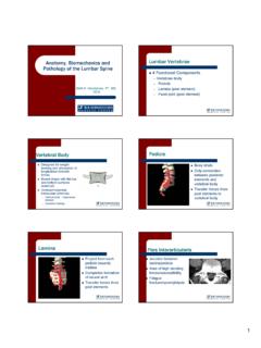
5 Diagnosis of lumbar radiculopathy is particularly challenging due to the anatomy involved. In the lumbar spine, the dorsal and ventral lumbar roots exit the spinal cord at the T11-L1 bony lev- Introduction el and travel in the lumbar canal as a group of nerve roots in the The dominant medical factors associated with the development dural sac known as the horse's tail' or cauda equina. Multiple of disability in Patients with low back pain is the presence of se- nerve rootlets that are descending in the cauda equina can be af- vere leg pain and a history of prior episodes of low back ,2 fected by a single central disk herniation or single level lumbar In Patients presenting with leg pain greater than low back pain, central stenosis.
6 For example, a central L3-4 disc herniations Clinical and Diagnostic Findings .. American Journal of Clinical Medicine Spring 2010 Volume Seven, Number Two 81. or central canal stenosis can impact the L5 and S1 nerve roots not acknowledged. The presenting symptoms and signs found This anatomy poses challenges to the diagnosis of upon examination of the distal lower extremities are similar be- lumbar radiculopathy and locating the compression site. tween polyneuropathy and lumbar radiculopathy. It is important that the practicing physician be able to recognize symptoms and The most useful test for confirming the presence of a radicu- signs that may be indicative of an overlying polyneuropathy lopathy is needle EMG (electromyogram).
7 An EMG study is with lumbar radiculopathy. In order to properly diagnose the considered Diagnostic for radiculopathy if muscles innervated co-existence of both disorders, imaging studies, and electrodi- by adjacent nerve roots are normal but EMG abnormalities are agnostic tests are needed. found in two or more muscles innervated by the same nerve root and different peripheral The needle EMG exami- The purpose of this study is to emphasize the importance of nation can identify only the root or roots that are physiologi- using Clinical symptoms and signs along with electrodiagnos- cally involved, not the precise anatomic site of pathology in tic and imaging studies to properly diagnose a polyneuropathy the lumbar spinal canal.
8 This is an important limitation which with radiculopathy. The frequency of polyneuropathy being di- requires correlation with imaging Findings to determine the ana- agnosed in Patients with lumbar radiculopathy who presented tomic location of the offending The most accurate imag- with low back and leg symptoms is studied. The researchers ing study to assess neural structures within the lumbar spine is review the common Clinical and Diagnostic Findings in these pa- MRI The needle EMG is helpful however, due to tients to provide practitioners with the identifiable combination the high false positive rate of lumbar spine MRIs with around of Clinical symptoms and signs that are most indicative of an 30% of normal subjects having a disk ,14 additional polyneuropathy.
9 The complexity of diagnosing this dual central and peripheral nerve lesion is acknowledged. Polyneuropathy is a common neurologic disorder affecting the peripheral nerves with a frequency among the general popula- tion above 5%.10 Pathophysiological changes can include: axo- Methods nal degeneration, axonal atrophy, demyelization, and metabolic Patients studied were all referred to a physician specialized changes that alter nerve ,16 Presenting symptoms in Electrodiagnostic Medicine and Physical Medicine and of polyneuropathy are described as pain, dysesthesias, and Rehabilitation for treatment of low back and lower extremity weakness in the feet and ,18 Signs and Findings associated symptoms.
10 The Patients seen with lumbar radiculopathy were with polyneuropathy are usually present with bilateral relative- counted. These Patients presented with low back pain and ra- ly symmetrical distal sensory loss, weakness, and hypoactive or dicular lower extremity symptoms of weakness, numbness, and absent ,3 The sensory loss is described to demonstrate pain. All these Patients had abnormal lumbar MRI Findings to a distal-to-proximal sensory loss gradient of small or large sen- confirm the diagnosis of lumbar radiculopathy. Of these pa- sory fibers. Signs of sensory loss occur in an acral, nondermato- tients, the ones clinically suspected and then diagnosed with mal, nonsingle-nerve distribution.