Transcription of 1 Compound microscope (Hund) 2 15 3 4 5 6 7 16 8 …
1 1) Eyepieces (magnifies 10x), one with diopteradjustment, 2) Interp[upillaryadjustment, 3) Head, 4) Revolving nosepiece, 5) Objectives (magnifies 4, 10, 40x, or 100x), 6) Slide holder, 7) Stage, 8) Iris diaphragm adjustment knob, 9) Light condenser, 10) Blue filter holder, 11) Condenser height adjustment knob, 12) Light source opening, 13) Field diaphragm adjustment, 14) Base, 15) Eyepiece diopteradjustment, 16) Stand, 17) Stage movement knobs, 18) Coarse focus knob, 19) Fine focus knob, 20) Light on/off switch & brightness151617181920123456789101112131 4 Compound microscope ( hund ) Compound Microscopes ) Eyepieces (magnifies 10x) diopter adjustment, 2) Interpupillaryadjustment, 3) Head, 4) Revolving nosepiece, 5) Objectives (magnify 4x, 10x, 20x, 40x, or 100x), 6) Slide holder, 7) Stage, 8) Iris diaphragm adjustment knob, 9) Light condenser, phase contrast see below, 10) Condenser height adjustment knob, 11) Field diaphragm adjustment, 12) Stand, 13) Light on/off switch, 14) Light intensity knob, 15) Stage movement knobs, 16) Fine focus knob, 17) Course focus knob, 18) BaseCompound microscope (Motic) Compound Microscopes Motic microscope (room 1660) is very similar to the hund microscope (room 1325) except that they are equipped with a phase contrast condenser.]
2 It is important the condenser turret be set at BFfor Bright Field or the number setting corresponding to the objective in use to get phase contrast effects. When done , please remove slide, return the turret to BF , and put the 4X objective back in Microscopes contrast works great for small cells or unstained or very thin sectionsCompound Microscopes Leave dust cover in cabinet Lift microscope with one hand on base, other on stand Gently set microscope down on counter Plug in, turn on and adjust light intensity about half way Open field and iris diaphragm about half way Put the red coded 4x objective in place (gives 40x magnification) Put slide on stage using clip holder Condenser should be just below stage (arrow on knob pointing up for Motics) Move stage using knobs so light shines through specimen Eyepieces diopter set at 0 for Motic or 65 for hund (line up number with white line) Set eyepiece distance between for your eyes Look though eyepiece and focus on specimen using course focus knob Turn revolving nose piece to yellow coded 10X objective to give 100x magn.
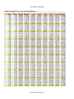
3 Use fine focus, turn to 40X objective (400x magn.) and fine focus as necessary Turn off light, unplug cord but please do NOT wrap it around microscope Put microscope back in same cabinet with the same dust cover onSet-up and careNote: This calibration was accomplished using a stage micrometer ruler 1 mm long divided into mm (= 10 m) markings mounted on a slide. * 20X objectives found on Motic scopes only and not all hund scopes have x1000 Step 1(same as when using the dissection microscope ): Measure two points on your drawing with the ruler from your student drawer. Make a horizontal line the same length beside the word Scale: . Step 2: Look at the actual specimen through the microscope and, using the ruler (ocular micrometer) in the right eyepiece, count the number of eyepiece divisions (epd) across same two points (this eyepiece ruler has no units -there are simply 100 eyepiece divisions on it). Multiply the epd by the corresponding number of micrometers given in the table below depending on which objective is in use to determine the width or length of the specimen in micrometers: ObjectiveMagnification1 epd=Example: view & draw a happy plant cell: Step 1: Draw a line beside the word Scale: the same width as you drewthe cell: Scale: Step 2: Measure the actualcell width using eyepiece ruler.
4 Multiply the number of eyepiece divisions by the number of micrometers that correspond to the objective in use using 10X objective: 4 epdx 10 m = 40 mIndicate scale beside drawing as: Scale: 40 m4Xx4025 m10Xx10010 m20X* mCompound Microscopes of scale using Compound microsco