Transcription of 12 Lead ECG Interpretation - Cleveland Clinic
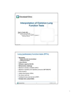
1 12 lead ecg interpretation Deborah Klein, MSN, RN, ACNS-BC, CCRN, CHFN, FAHA. Clinical Nurse Specialist, Coronary ICU, Heart Failure ICU, Cardiac Short Stay/PACU/CARU. Nursing Institute 1. Confidential DOSO xtober Course 2010. 2017. Cleveland Clinic 2017. 12 Lead ECG (or EKG). Heart is an electrical field; arms and legs are a linear extension of this field ECG is a recording of the electrical activity of the heart over a period of time Detected by electrodes attached to the surface of the skin and recorded and displayed by a device external to the body Changes in electrical activity may indicate arrhythmias, cardiac ischemia, or electrolyte imbalances 2 DOS Course 2017. 3 DOS Course 2017. 4 DOS Course 2017. Blood Supply LAD: anterior wall of LV, anterior septum, bundle branches Left circumflex: left atrium, lateral wall LV, posterior wall LV.
2 RCA: right atrium, right ventricle, bottom of LV, posterior septum 5 DOS Course 2017. 12 Lead ECG System 3 limb leads (bipolar). 3 augmented limb leads (unipolar). 6 precordial leads 6 DOS Course 2017. Limb Leads: Bipolar Leads I, II, and III. Two electrodes (+ and -) equidistant from heart Records electricity flow from negative to positive electrode A wave of depolarization moving toward a positive electrode produces a positive deflection on the ECG. Depolarization moving away from a positive electrode records a negative deflection Lead axis is the direction of electrical depolarization 7 DOS Course 2017. 8 DOS Course 2017. Limb Leads: Unipolar Leads aVR, aVL, aVF. Letter a refers to augmented Letter V refers to voltage Letters R, L, and F refer to where positive electrode is placed (right arm, left arm and left leg).
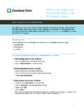
3 Records electricity flow from center of heart toward positive electrodes 9 DOS Course 2017. 10 DOS Course 2017. 11 DOS Course 2017. Limb Lead Placement 12 DOS Course 2017. Limb Lead Electrode Placement Preferred site of limb lead electrodes is slightly proximal to wrist and ankles over flat fleshy area Upper arms and legs may be used but must be consistent Avoid muscle and boney areas 13 DOS Course 2017. Precordial Leads 6 precordial leads (V1 V6). Letter V refers to unipolar Numbers 1-6 are codes for locations on precordium 14 DOS Course 2017. Precordial Leads V1 and V2 are on either side of sternum at 4th ICS. V4 is midclavicular line, 5th ICS. V3 is halfway between V2 and V4. V6 is at midaxillary line, 5th ICS. V5 is halfway between V4 and V6, 5th ICS. 15 DOS Course 2017. 16 DOS Course 2017. Precordial Lead Electrode Placement Correct anatomical placement imperative!
4 Sternal angle (angle of Louis) used as reference point Run finger down the sternum, from the sternal notch at the top until a boney horizontal ridge, the sternal angle is met With your finger on this ridge, slide down and to the right side to locate the second intercostal space Count down to the third and fourth space Locate the edge of the sternum and place V1. In women, V4, V5, and V6 are place under the left breast 17 DOS Course 2017. View from Precordial Leads V1 Right ventricle V2/V3 Septum V4 Apex V5/V6 LV; left lateral wall 18 DOS Course 2017. 19 DOS Course 2017. V6. 0 . V5. 30 . V1 V4. 120 V3 60 . V2. 75 . 90 . 20 DOS Course 2017. Skin Preparation Assess the skin If visibly oily or sweaty prepare the skin before electrode placement Cleanse the sites for electrode placement using the following options: Soap and water and dry thoroughly Alcohol and gauze pads Abrading the skin to remove dead skin with a washcloth Clip hair for a 2x2 area for each electrode if necessary 21 DOS Course 2017.
5 12 Lead ECG Lead Wires Lead wires fasten to the electrodes 22 DOS Course 2017. 12 Lead ECG Quality All tracings must have a clean stable baseline free of artifact and interference Artifact can be caused by: Muscle tremors Patient movement Loose electrodes 60 cycle interference 23 DOS Course 2017. Poor Quality Wandering Baseline 24 DOS Course 2017. Good Quality 25 DOS Course 2017. Poor Quality 26 DOS Course 2017. Goal is Quality Tracings! Proper placement = accurate tracings = accurate treatment = quality patient outcomes 27 DOS Course 2017. 29 DOS Course 2017. 30 DOS Course 2017. Basic 12 lead ecg interpretation Determine rate Bradycardia, less than 60 beats per minute Tachycardia, greater than 100 beats per minute Determine rhythm Regular or irregular Ischemia, injury or infarction? 31 DOS Course 2017. 32 DOS Course 2017.
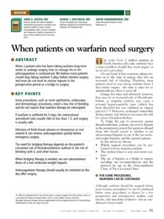
6 Definitions Ischemia 70% of vessel occluded O2 demand exceeds supply Injury Ischemic state continues with injury to myocardium Infarction Cell death Ischemia and injury are reversible Infarction is not reversible 34 DOS Course 2017. Ischemia T wave inversion; symmetric, narrow ST depression of 1-2 mm or more for a duration of seconds in the leads facing the ischemic area Reversible 35 DOS Course 2017. 36 DOS Course 2017. Injury ST elevation over damaged myocardium Downward concave or coned shape Merge with T wave Reversible 37 DOS Course 2017. 38 DOS Course 2017. Infarction Irreversible Seen on ECG in stages Hyperacute Tall, narrow, peaked T waves Invert within a few hours 39 DOS Course 2017. Infarction ST segment elevation Seen in early hours of infarction Last from several hours to several days Reciprocal changes: leads facing away from infarction may show ST depression Q waves seconds or more wide 1/4 to 1/3 height of R wave Develop within several hours to 48 hours after infarction 40 DOS Course 2017.
7 41 DOS Course 2017. Inferior Wall MI. Supplied by RCA. Leads II, III, and aVF. Damage may extend into RV. Biventricular dysfunction SA node dysfunction Bradyarrhythmias Heart blocks 42 DOS Course 2017. Anterior Wall MI. Supplied by LAD. Changes in precordial leads V1-V6. ST elevation Q waves Arrhythmias Ventricular (PVCs). Bundle branch blocks 44 DOS Course 2017. Lateral Wall MI. Supplied by left circumflex artery Leads I, aVL, V5 and V6. Potential for reduction in LV function, but not as great as with anteroseptal wall MI. Arrhythmias from SA node dysfunction Sinus arrest Bradyarrhythmias 46 DOS Course 2017. Posterior Wall MI. Reciprocal changes Tall R waves ST segment depression Look in leads opposite posterior wall (V1, V2). Frequently seen with inferior wall MI. Leads II, III, and aVF. Junctional rhythm, heart blocks 47 DOS Course 2017.
8 Pericarditis Signs and symptoms mimic an MI. Diagnosis based on clinical presentation, 12 lead ECG, and echocardiogram Clinical Presentation Sharp, pleuritic chest pain Worse on inspiration Pain relieved by sitting up or leaning forward No response to NTG. Pericardial rub 49 DOS Course 2017. 12 Lead ECG Findings Diffuse changes that may not localize to right or left coronary artery distribution Diffuse ST elevation in multiple leads PR segment depression Sinus tachycardia or atrial arrhythmias 50 DOS Course 2017. Mrs. T. 62 year old woman with a history of rheumatic fever, panic attacks, tobacco use (1/2 pack/day for 8 years). Last evening while walking up the stairs she had right sided chest pain radiating to the neck and over her sternum. Pain was a pressure sensation at 10/10 at its worst Diaphoresis and chills with the chest pain Last week she noticed worsening chest pain with exertion when walking to the bus or going up the stairs Pain subsided with rest On arrival to ED: HR 74 bpm, BP 196/61 mmHg After one SL nitroglycerine, BP 160/79 mmHg 52 DOS Course 2017.
9 54 DOS Course 2017. Mrs. M. 72 year old woman complaining of sudden onset of midepigastric pain and diaphoresis Past medical history Atrial fibrillation Known CAD with stent placement to the RCA in 2009. Former smoker (quit 1/1/2003). Medications Dofetilide (tikosyn), metoprolol ASA, ticagrelor (brilinta), atorvastatin (lipitor). 55 DOS Course 2017. Mr. G. 29 year old male with a history of substance abuse At 4 pm on 1/05 he injected a combination of heroin and cocaine He was found unresponsive by his father who called EMS. EMS gave him received 3 doses intranasal naloxone (Narcan) with no response; he was transported to the ED. In the ED he received 2 mg naloxone (Narcan) through an intraosseous (IO) device and became responsive He complained of 10/10 chest pain 58 DOS Course 2017. 61 DOS Course 2017.