Transcription of 13.24: Mass Spectrometry - Vanderbilt University
1 1 Chapter 13: spectroscopy Methods of structure determination Nuclear Magnetic Resonances (NMR) spectroscopy (Sections ) Infrared (IR) spectroscopy (Sections ) ultraviolet - visible (UV-Vis) spectroscopy (Section ) Mass (MS) Spectrometry (not really spectroscopy ) (Section ) Molecular spectroscopy : the interaction of electromagnetic radiation (light) with matter (organic compounds). This interaction gives specific structural information. 2 : Mass Spectrometry : molecular weight of the sample formula The mass spectrometer gives the mass to charge ratio (m/z), therefore the sample (analyte) must be an ion. Mass Spectrometry is a gas phase technique- the sample must be vaporized. Electron-impact ionization Sample Inlet10-7 - 10-8 torrR-Helectr on beam 70 eV (6 700 KJ/mol)e_R-H+massanalyze rm/zioniza tion chamber(M+)proton u neutron u electron u 13 mass m charge z ==B2 r2 2V B= magnetic field strength r = radius of the analyzer tube V= voltage (accelerator plate) The Mass Spectrometer Io niz atio nchamberIo ns of selectedmas s/charge rat ioare detect edIo ns o f n on-selectedmass/char ge r at ioar e not detect edMagnetic Field, Bo 4 Exact Masses of Common Natural Isotopes Isotope mass natural abundance 1H 2H 12C 13C ( ) 14N 15N ( ) 16O 17O ( ) 18O ( ) Isotope mass natural abundance 19F 35Cl 37Cl ( ) 79Br 81Br (98%) 127I Dalton (Da) or mass unit (u)
2 = units for measuring molecular masses. One Da. = 1/12 the mass of the 12C atom Monoisotopic (exact) mass sum of the exact masses of the most abundant isotope of each element in a molecule Average mass sum of the averaged masses of each element in a molecules, weighted according to isotopic abundance. Nominal mass mass calculated using the integer mass of the most abundant isotope for each element (H=1, C=12, O=16, N=14, etc.) 25 Molecular Ion (parent ion, M) = molecular mass of the analyte; sample minus an electron. (mass of an e is 1/1836 that of a proton) Base peak- largest (most abundant) peak in a mass spectrum; arbitrarily assigned a relative abundance of 100%. C6H6m/z = (M+) (100%) m/z=79 (M+1) (~ of M+) 6 The radical cation (M+ ) is unstable and will fragment into smaller ions Relative abundance (%) m/z=16 (M+) m/z=15 m/z=14 m/z=17 (M+1) CHHH+H+m/z = 15cha rge neutralnot detec tedCH+HHcharge neutr alnot detecte dm/z = 1 4 CHHHH- e_CHHHH+m/z = 16+Relative abundance (%) m/z m/z m/z=15 m/z=29 m/z=43 m/z=45 (M+1) m/z=44 (M) CHHCCHHHHHHCHHCCHHHHHH+CHHCCHHHHH+H+char ge ne utral notdet ec tedm/z = 43m/z = 4 4 CHHCCHHHHHH- e_+CHHCHHH+CHHH+CHHCHHH+CHHH+m/z = 2 9m/z = 15charg e ne utra lnot detectedcha rge ne utralnot detec ted- e_37 m/z m/z ClBrm/z=112 (M+) m/z=113 (M+ +1) m/z=114 (M+ +2) m/z=115 (M+ +3) m/z=77 35Cl 37Cl ( ) 79Br 81Br (98%) m/z=77 m/z=156 (M+) m/z=158 (M+ +2) m/z=157 (M+ +1) m/z=159 (M+ +3) Mass spectrum of halogenated organic compounds 8 Mass spectra can be quite complicated and interpretation difficult.
3 Some functional groups have characteristic fragmentations It is difficult to assign an entire structure based only on the mass spectrum. However, the mass spectrum gives the mass and formula of the sample, which is very important information. To obtain the formula, the molecular ion must be observed. (Soft ionization techniques) Methods have been developed to get large molecules such as polymers and biological macromolecules (proteins, peptides, nucleic acids) into the vapor phase 49 : Molecular Formula as a Clue to Structure Nitrogen rule: In general, small organic molecules with an odd mass must have an odd number of nitrogens. Organic molecules with an even mass have zero or an even number of nitrogens. If the mass can be determined accurately enough, then the molecular formula can be determined (high-resolution mass Spectrometry ). Information can be obtained from the molecular formula: Degrees of unsaturation: the number of rings and/or -bonds in a molecule (Index of Hydrogen Deficiency).
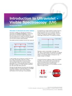
4 10 Degrees of unsaturation saturated hydrocarbon CnH2n+2 cycloalkane (1 ring) CnH2n alkene (1 -bond) CnH2n alkyne (2 -bonds) CnH2n-2 For each ring or -bond, 2H from the formula of the saturated alkane HHHHHHHHHHHH C6H14 - C6H12 H2 2 = 1 1 2 C6H14 - C6H6 H8 8 = 4 1 2 HHHHHHH ydrogen Deficiency Degrees of Unsaturation 511 Correction for other elements: For Group VII elements (halogens): subtract 1H from the H-deficiency for each halogen, For Group VI elements (O and S): No correction is needed For Group V elements (N and P): add 1H to the H-deficiency for each N or P C12H4O2Cl4 C10H14N2 12 : Principles of molecular spectroscopy : Electromagnetic radiation = distance of one wave = frequency: waves per unit time (sec-1, Hz) c = speed of light ( x 108 m sec-1) h = Plank s constant ( x 10-34 J sec) Electromagnetic radiation has the properties of a particle (photon) and a wave.
5 Organic molecule (ground state) light h organic molecule (excited state) organic molecule (ground state) + h relaxation 613 h c Quantum: the energy of a photon E = h = E = c (cm) short high high Wavelength ( ) Frequency ( ) Energy (E) long low low E E -1 -1 1010-2radiowavesmicrowaves10-4infraredVi s10310-7 UltravioletX-Rays Rays10-1110-510-914 : Principles of Molecular spectroscopy : Quantized Energy Levels molecules have discrete energy levels (no continuum between levels) A molecule absorbs electromagnetic radiation when the energy of photon corresponds to the difference in energy between two states 715 UV-Vis: valance electron transitions - gives information about -bonds and conjugated systems Infrared: molecular vibrations (stretches, bends) - identify functional groups Radiowaves: nuclear spin in a magnetic field (NMR) - gives a map of the H and C framework organic molecule (ground state) light h organic molecule (excited state) organic molecule (ground state) + h relaxation 16 ultraviolet - visible (UV-Vis) spectroscopy 200 UV 400 800 nm Vis Recall bonding of a -bond 817 -molecular orbitals of butadiene 1: 0 Nodes 3 bonding interactions 0 antibonding interactions BONDING MO 2: 1 Nodes 2 bonding interactions 1 antibonding interactions BONDING MO 3: 2 Nodes 1 bonding interactions 2 antibonding interactions ANTIBONDING MO 4: 3 Nodes 0 bonding interactions 3 antibonding interactions ANTIBONDING MO 2 is the Highest Occupied Molecular Orbital (HOMO) 3 is the Lowest Unoccupied Molecular Orbital (LUMO) 18 UV-Vis light causes electrons in lower energy molecular orbitals to be promoted to higher energy molecular orbitals.
6 HOMO LUMO Chromophore: light absorbing portion of a molecule But adieneBut adiene919 Molecular orbitals of conjugated polyenes BondingAntibondingEnergyH2 CCH2180 nm217 nm258 nm290 nm20 violet 400 nm yellow blue 450 orange blue-green 500 red yellow-green 530 red-violet yellow 550 violet orange 600 blue-green red 700 green Color of absorbed light Color observed 380 nm780 nm400 nm450 nm500 nm550 nm600 nm700 nmredorangeyellowgreenblueviolet-indigoM olecules with extended conjugation move toward the visible region 1021 NNNNMgOCO2CH3 OOChlorophyll -carotene lycopene OHOOHOHOOHOHOHOOHOHOH+anthocyanin Many natural pigments have conjugated systems 22 Chromophore: light absorbing portion of a molecule Beer s Law: There is a linear relationship between absorbance and concentration A = c l A = absorbance c = concentration (M, mol/L) l = sample path length (cm) = molar absorptivity (extinction coefficient) a proportionality constant for a specific absorbance of an analyte 1123 : Introduction to Infrared spectroscopy E 1 is expressed as (wavenumber), reciprocal cm (cm-1) _ = 1 E _ therefore _ (cm) VisNearIRFarIRInfrared (IR)micr x 10-4 x 10⁻³ cm16 m10-410-2 _4000 _600IR radiation causes changes in a molecular vibrations 24 Symmetric stretch Antisymmetric stretch Stretch: change in bond length in-plane bend out-of-plane bend scissoring rocking wagging twisting Bend: change in bond angle >>>>>>>>>>>>>>>>Animation of bond streches and bends: 1225 Bond Stretch.
7 Hooke s Law =_2 c1mx mymx + myf12XY = vi bra tional frequencyc = sp eed of lightmx = mass of Xmy = mass of Y_mx mymx + my= reduce d mass ( )f = sp ring co nst ant; typ e of bond between X and Y (si ngle, double or tri ple)E f _ Hooke s law simulation: 26 Infrared Spectra Interpretation of an Infrared Spectra: Organic molecules contain many atoms. As a result, there are many stretching and bending modes and the IR spectrum has many absorption bands Four distinct regions of an IR spectra 40 00 cm-160 0 cm-115 00 cm-1fingerprintregiondoublebondre gion20 00 cm-125 00 cm-1tri plebondregionX-Hsi ngle bondre gionC-H O-H N-H C C C N C=C C=O 500 cm-1 1327 Fingerprint region (500 - 1500 cm-1): low energy single bond stretching and bending modes. The fingerprint region is unique for any given organic compound. However, there are few diagnostic absorptions. Double-bond regions (1600 - 1900 cm-1) C=C 1620 - 1680 cm-1 C=O 1680 - 1850 cm-1 Triple-bond region.
8 (2100 - 2300 cm-1) C C 2100 - 2200 cm-1 (weak, often not observed) C N 2240 - 2280 cm-1 X-H Single-bond region (2800 - 3600 cm-1) O-H 3200 - 3600 cm-1 (broad) CO-OH 2500 - 3600 cm-1 (very broad) N-H 3350 - 3500 cm-1 C-H 2800 - 3300 cm-1 sp3 C-H 2850 - 2950 cm-1 sp2 =C-H 3000 - 3100 cm-1 sp C-H 3310 - 3320 cm-1 28 Alkenes =C-H 3000 - 3100 cm-1 medium - strong C=C 1620 - 1680 cm-1 medium Aromatic =C-H 3000 - 3100 cm-1 strong C=C 1450 - 1600 cm-1 strong Alkynes C-H 3310 - 3320 cm-1 strong C C 2100 - 2200 cm-1 weak - medium Alcohols C-O 1025 - 1200 cm-1 strong O-H 3200 - 3600 cm-1 strong and broad Amines C-N 1030 - 1230 cm-1 medium N-H 3350 - 3500 cm-1 medium Carbonyl C=O 1680 - 1850 cm-1 strong Carboxylic acids O-H 2500 - 3500 cm-1 strong and very broad Nitrile C N 2240 - 2280 cm-1 weak-medium Characteristic Absorption Frequencies Ta b l e 1 3 . 3 , p . 5 5 2 1429 cm-1 C-H C-H C C CCHH3CH2CH2CH2C% transmittance % transmittance cm-1 C-H C C CCCH3H3CH2CH2CC-H hexane cm-1 % transmittance C-H C=C =C-H % transmittance cm-1 H3CH2CH2CH2 CHCCHH% transmittance cm-1 C=C C-H =C-H CCH3CH3CH2CH2 CHH30 O-H C-H C-O CH3(CH2)4CH2OH CH3(CH2)4CH2NH2 N-H C-H CH3(CH2)4CH2NH-CH3 N-H C-H C-H C N H3C(H2C)4CH2C N % transmittance % transmittance 1531 OCOCH2CH3H3C(H2C)3H2 COCOH(H3C)3CH2CO-H C-H C=O 1730 cm-1 OCHH3C(H2C)3H2CC-H OCCH3H3C(H2C)2H2CC=O 1715 cm-1 C=O 1710 cm-1 C=O 1705 cm-1 OCH% transmittance % transmittance 32 40006003000200010004000600300020001000X- H Regon(2500-4000 cm-1)C-HO-HN-HTriple BondRegion(2000-2500)CCCND ouble BondRegion (1500-2000)Fingerprint Region(600-1500 cm-1)COCNCC difficult to interpretAlkanes, Alkyl groups C-HAlkenes =C-H C=CAlkynes C-H C CAlkyl Halides C-Cl C-Br C-IAlcohols O-H C OAromatics =C-H2850-2950 (m-s)3000-3100 (m)1620-1680 (m-w)3310-3320 (s)
9 2100-2260 (m-w)600-800500-6005003200-3600 (br, m-s)1025-1200 (s)3000-3100 (m-w)1450-1600 (m-w)Amines N-HAmide N-H C=OCarbonyl Group C=O C OCarboxylic Acids O-H C=ONitriles C NNitro Group -NO23350-3500 (br, m)3180-3350 (br, m-w)1680-1700 (s)1650-1780 (s)~1200 (s)2500-3100 (br, s)1700-1725 (s)2240-2250 (w-m)1540cm-1 Functional Groupcm-1 Typical IR Absorptions for Funtional GroupsFunctional Group1633 : Introduction to 1H NMR direct observation of the H s of a molecules Nuclei are positively charged and spin on an axis; they create a tiny magnetic field ++Not all nuclei are suitable for NMR. 1H and 13C are the most important NMR active nuclei in organic chemistry Natural Abundance 1H 13C 12C (not NMR active) 34 (a) Normally the nuclear magnetic fields are randomly oriented (b) When placed in an external magnetic field (Bo), the nuclear magnetic field will either aligned with (lower energy) or oppose (higher energy) the external magnetic field Fig , p.
10 513 1735 The energy difference between aligned and opposed to the external magnetic field (Bo) is generally small and is dependent upon Bo Applied EM radiation (radio waves) causes the spin to flip and the nuclei are said to be in resonance with Bo E = h E = Bo h 2 Bo = external magnetic field strength = gyromagnetic ratio 1H= 13C= Note that is a constant and is sometimes denoted as h h 2 ++++36 NMR Active Nuclei: nuclear spin quantum number (I) atomic mass and atomic number Number of spin states = 2I + 1 (number of possible energy levels) Even mass nuclei that have even number of neutrons have I = 0 (NMR inactive) Even mass nuclei that have an odd number of neutrons have an integer spin quantum number (I = 1, 2, 3, etc) Odd mass nuclei have half-integer spin quantum number (I = 1/2, 3/2, 5/2, etc) I= 1/2: 1H, 13C, 19F, 31P I= 1: 2H, 14N I= 3/2: 15N I= 0: 12C, 16O 1837 Continuous wave (CW) NMR Pulsed (FT) NMR Fig.