Transcription of 145 Interpretation of the Chest Radiograph - Part 1
1 Sign up to receive ATOTW weekly - email ATOTW 145 Interpretation of the Chest Radiograph - part 1, 10/08/09 Page 1 of 10 Interpretation OF THE Chest Radiograph PART 1. ANAESTHESIA TUTORIAL OF THE WEEK 146 10TH AUGUST 2009 Dr. Tim Dawes Royal Devon and Exeter Hospital, UK Dr. Gerry Lynch Rotherham General Hospital, UK Correspondence to PART 1: TECHNICAL ASPECTS, A STANDARD Interpretation . ROUTINE AND COMMON ABNORMALITIES OF THE Chest Radiograph This is part 1 of a 3-part series of tutorials. Parts 2 and 3 will focus more specifically on the Chest Radiograph changes commonly seen on the intensive care unit.
2 MULTIPLE CHOICE QUESTIONS 1. Concerning the normal Chest Radiograph : a. The right mediastinal border is formed by the right brachiocephalic vein, SVC and right hilum b. The right hilum lies 1-2 cm above the left c. The horizontal fissure runs from the hilum to the 6th rib in the axillary line d. The right ventricular border is visible on the frontal film 2. Concerning the frontal Chest Radiograph : a. The medial heads of the clavicles should be equidistant from the tracheal midpoint in an unrotated film b. An adequate breath should result in 5 rib ends visible anteriorly c.
3 Sternal fractures are readily visible d. Obscuration of the right heart border indicates right lower lobe disease 3. Concerning the frontal Chest Radiograph : a. The cardiothoracic ratio should be less than 1:2 on the PA film b. This ratio is decreased in expiration c. The lingula is considered to form part of the left lower lobe d. The oblique fissure is visible on the frontal film Sign up to receive ATOTW weekly - email ATOTW 145 Interpretation of the Chest Radiograph - part 1, 10/08/09 Page 2 of 10 INTRODUCTION The Chest Radiograph , or X-ray (CXR) is one of the commonest investigations requested for intensive care patients.
4 Its correct Interpretation is therefore an integral skill for ICU clinicians. As with history and examination skills, a structured approach is necessary to avoid missing important signs. Interpretation of the CXR does not take the place of a good history and examination, but should be seen as an extension of the examination process. A firm grasp of the normal Chest film is an essential start point. This article does not aim to cover all aspects of CXR Interpretation , rather to give an introduction to Interpretation and highlight aspects which are particular to intensive care patients.
5 TECHNICAL ASPECTS The Chest is imaged by positioning the patient between an X-ray source and an X-ray sensitive plate. The distance from the source to the patient is relatively great compared to the distance from the patient to the plate in order to reduce magnification of the image and problems of rotation of the structures within the patient. Even so, the structures nearest the plate will have their size most faithfully represented, whereas those furthest from the plate will be subject to a small but significant degree of magnification.
6 The patient may be orientated in several ways: anterior-posterior ( AP with the patient facing the source, standard in ICU, lower radiation dose, quality affected by rotation) posterior-anterior ( PA with the patient facing the plate, higher xray dose, less affected by rotation) or lateral, expiratory, lordotic or decubitus This discussion will be confined to AP and PA films since these are by far the most common on intensive care. The term frontal view will be used to represent PA or AP collectively in contrast to lateral views.
7 Different tissues absorb X-rays to different extents, resulting in a spectrum of exposures onto the plate and a corresponding spectrum of shades of grey on the final film. High density (radio-opaque) tissues such as bone and metal ( prosthetic heart valves, coronary stents) appear white. Low-density (radiolucent) tissues such as air appear black. Fluid ( blood, consolidation) and fat are moderately absorbent and therefore appear grey. Standard Assessment Routine Interpretation of the CXR can be considered in three stages: preliminaries, the film itself, and synthesis of these abnormalities into a differential diagnosis 1.
8 Preliminaries It is common for students to be asked to interpret a CXR in front of peers or seniors only seconds after it has been brandished in front of their eyes. A careful introduction to the film in question is not only important to avoid assigning abnormalities to the wrong patient, but also allows a few extra seconds to scan the film. Check the following: Name Is the Radiograph from the patient you re interested in discussing? Date Is the date of the CXR the date you were expecting? Intensive care patients typically accrue several CXRs on their journey to the ICU, and may even have films from when they were clinically well Sign up to receive ATOTW weekly - email ATOTW Interpretation of the CXR in the ICU 1, Page 3 of 10 Orientation Is this an AP or a PA film look for this written on the film, or the comment supine suggesting an AP film.
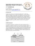
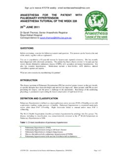
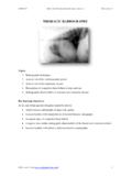
9 Is the film displayed the correct way around? Look for the L or R markers added to the film. More cruel examiners have been known to present a CXR of a patient with dextrocardia the wrong way around to see if students check the orientation markers placed by the radiographer Figure 1. CXR of a patient with dextrocardia. Note the white orientation tab in the top left of the film ( R ) confirming that the film has been displayed the correct way around. This patient also has a left sided pleural effusion (e) Adequacy Can you see the area you need to see?
10 Check the lung apices are visible. If the CXR was indicated for naso-gastric tube placement then check it is visible. Ideally an adequate breath allows five ribs to be visible anteriorly. Rotation The medial ends of the clavicles should be equidistant from the spinous processes that project between the clavicular heads. This is shown in figure 2 in the upper inset. The dimension A equals the dimension B. Penetration Three lower thoracic vertebrae and the pulmonary vessels to the left lower lobe should be visible through the cardiac silhouette.