Transcription of 163 Management of subarachnoid haemorrhage - …
1 Sign up to receive ATOTW weekly - email ATOTW 163. Management of subarachnoid haemorrhage , 07/12/2009 Page 1 of 11 MANAGEMEMT OF subarachnoid haemorrhage ANAESTHESIA TUTORIAL OF THE WEEK 163 7th DECEMBER 2009 Sarah Davies, Locum Consultant Anaesthetist Leeds General Infirmary, Leeds, UK Correspondence to QUESTIONS 1. The following are risk factors for a poor outcome post aneurysmal subarachnoid haemorrhage (SAH): a. Male sex b. Presence of co-morbid conditions c. Anterior circulation aneurysm d. World Federation of Neurosurgeons (WFNS) grade IV 2. Proven therapies in the Management of vasospasm include: a. Triple H therapy b. Oral nimodipine c. Antifibrinolytics d. Balloon angioplasty 3. The following statements are true: a. The ISAT study demonstrated a better outcome for coiled versus clipped aneurysms b. The overall case mortality for SAH is 10% c. Intraoperative hypothermia improves neurological outcome d. CT is 100% sensitive in detecting aneurysms INTRODUCTION subarachnoid haemorrhage (SAH) is a type of stroke, which occurs when there is bleeding in the subarachnoid space around the brain.
2 The incidence in the UK is approximately 8 per 100,000 population per year ( ). The commonest cause of SAH is rupture of a cerebral aneurysm (70-85%) - other causes include arteriovenous malformations, tumours, trauma and non-aneurysmal perimesencephalic haemorrhage . This tutorial addresses the Management of spontaneous aneurysmal SAH. The anaesthetist may be involved in the Management of patients with SAH at several stages; initial resuscitation and stabilisation, therapeutic Management in the ICU and providing anaesthesia perioperatively for aneurysmal occlusion. The prognosis is poor, with an overall case fatality of 50%, however early treatment has been shown to improve outcome, and it is therefore imperative that the anaesthetist is familiar with all aspects of the condition, and up to date with the latest evidence based Management . The mainstays of treatment are occlusion of the aneurysm, by endovascular coiling or surgical clipping, and prevention of cerebral ischaemia.
3 Sign up to receive ATOTW weekly - email ATOTW 163. Management of subarachnoid haemorrhage , 07/12/2009 Page 2 of 11 EPIDEMIOLOGY/AETIOLOGY SAH accounts for 5-15% of all strokes, and has an overall case fatality of around 50% with a trend towards gradual improvement. This includes the 10-15% of patients who die before reaching hospital, the rest dying (almost always) within the first 3 weeks, secondary to re-bleeding and/or cerebral vasospasm. Of those who survive, one third remain moderately to severely disabled. The rate of re-bleed is 2-4% within the first 24 hours, and 15-20% within the first 2 weeks if left un-occluded. The main predictors of mortality and dependence are impaired level of consciousness on presentation, advanced age and a large volume of blood on initial CT scan. The National Study of SAH described patient characteristics and aneurysm aetiology, based on findings from 2397 cases.
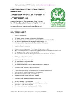
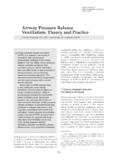
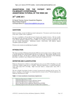
4 The median age of patients was 52 years, of which 66% were women. The majority of aneurysms (89%) were located in the anterior (carotid) circulation, the rest being in the posterior (vertebrobasilar) circulation. In 25% of cases aneurysms are multiple. Almost half of the patients had concurrent medical conditions such as hypertension and ischaemic heart disease. PATHOPHYSIOLOGY Intracranial aneurysms are most likely to develop over the course of an individual s life, with only 10% accountable to a genetic/familial cause. Modifiable risk factors include hypertension, smoking and excessive alcohol intake, all of which approximately double the risk of aneurysmal development. Aneurysms develop mostly where there is turbulent flow at vascular bifurcations, on or near to the Circle of Willis. The most common sites are illustrated in Figure 1. Only a small proportion is attributable to infection or trauma.
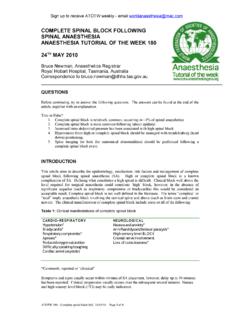
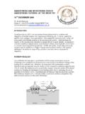
5 The majority are small, between 5-10 mm, when they rupture. Figure 1: Common sites of aneurysms within the cerebral circulation. ACA - anterior cerebral artery; A com anterior communicating artery; ICA internal carotid artery; P comm. posterior communicating artery; BA basilar artery. Sign up to receive ATOTW weekly - email ATOTW 163. Management of subarachnoid haemorrhage , 07/12/2009 Page 3 of 11 Figure 2: A giant left carotid artery aneurysm (arrow) Figure 3: A giant left hemisphere arteriovenous malformation (AVM) CLINICAL PRESENTATION The most characteristic presenting symptom is a sudden onset severe headache, accounted for by the sudden rise in ICP which occurs at rupture. This is often accompanied by the development of meningism (meningeal irritation caused by blood in subarachnoid space), seizures, confusion/agitation and focal neurological deficits (due to focal ischaemia and/or cranial nerve involvement).
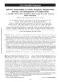
6 By the time of admission, two thirds of patients have a depressed level of consciousness. A GCS score of less then 8 is usually associated with increased ICP. Systemic dysfunction is common in the acute phase, and may include cardiac ischaemia, and neurogenic pulmonary oedema. There are several grading systems based on both clinical assessment and investigations. The World Federation of Neurosurgeons (WFNS) clinical grading system is the most widely used in the UK. (Table 1). The Fischer scale is described in Diagnosis. Table 1: WFNS Grading scale for SAH. GCS, Glasgow Coma Score. Grade GCS Score Motor Deficit I 15 Absent II 13 or 14 Absent III 13 or 14 Present IV 7 12 Either V 3 6 Either DIAGNOSIS In the majority of cases the diagnosis of SAH is made by unenhanced (non-contrast) CT (Figure 3). Cranial CT is indicated in all patients complaining of sudden onset severe headache lasting for longer than one hour and with no alternative explanation.
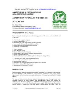
7 It should be performed as soon as possible after the patient presents, since SAH may not be evident on delayed CT if blood reabsorption has begun. CT may miss 2-7% of subtle SAH and in the case of a negative CT, further investigations are required. These include lumbar puncture, angiography (Figure 5B and 5C) and MRI or MRA (magnetic resonance angiography). The amount of blood on CT can be described by the Fischer grading scale, which is scored from 1 to 4, 4 being the most extensive. It may be the best predictor of cerebral vasospasm complicating SAH and overall patient outcome. The location and distribution of blood on a positive CT aids the identification of the cause of the SAH, be it an aneurysm or other pathology. There are three methods to delineate this; contrast CT angiography, magnetic resonance angiography and catheter angiography, which is the gold standard. Angiography not only indentifies one (or more) aneurysm, it details the anatomical configuration, which allows optimum selection of occlusion Management (coiling or clipping).
8 The most recent development is in 3-D rotational angiography, which gives more detailed assessment of the aneurysm (Figure 5C). Sign up to receive ATOTW weekly - email ATOTW 163. Management of subarachnoid haemorrhage , 07/12/2009 Page 4 of 11 Figure 4: A non-contrast CT scan showing a large amount of subarachnoid blood A B C Figure 5: Imaging of a right 3cm diameter posterior communicating artery aneurysm. A, non-contrast CT; B, angiogram via right carotid artery; C, digital reconstruction of aneurysm to assess suitability for coiling Management There are 3 major neurological complications post ruptured cerebral aneurysm: Re-bleeding (Figure 6A), cerebral vasospasm leading to ischaemia (Figure 6B), and hydrocephalus (Figure 6C). A B C Figure 6: A, Right hemisphere bleed after clipping of an aneurysm; B,Left hemisphere infarction complicating vasospasm after SAH; C, Hydrocephalus post-SAH Sign up to receive ATOTW weekly - email ATOTW 163.
9 Management of subarachnoid haemorrhage , 07/12/2009 Page 5 of 11 Additionally, there are several systemic manifestations, most importantly, cardiopulmonary dysfunction and electrolyte disturbances. Management is therefore targeted at prevention of rebleeding by means of aneurysm occlusion, and Management of complications. Depending on the neurological state of the patient, they will need to be managed on either the High Dependency Unit, or intubated and ventilated on Intensive Care. General neuroprotective strategies should be employed on ICU, particularly; adequate sedation, control of oxygenation and ventilation (CO2 levels), avoidance of hypotension, prevention of hyperthermia, and normoglycaemia. Occlusion Therapy This can be done either surgically (clipping at craniotomy) or using a radiological endovascular technique with detachable coils (coiling). Early occlusion prevents re-bleeding.
10 Evacuation of subarachnoid blood (at clipping) may also reduce the incidence of vasospasm. Early clipping or coiling, within 72 hours, is now the goal for all grades of SAH (WFNS I -V), replacing the out-dated approach of delayed aneurysm occlusion (until after the period of vasospasm) for poor grade SAH. Figure 7: Angiogram showing aneurysm post-coiling To clip or to coil? Neurosurgical aneurysm clipping requires a craniotomy, performed under general anaesthesia. It takes 4 8 hours, and has a procedural mortality rate of 1-3%. Coiling is a minimally invasive percutaneous endovascular treatment, which has proved to be a safe alternative to traditional surgical clipping of the aneurysm, and may be associated with a better outcome in selected patients. The technique consists of packing the aneurysm with detachable coils, and is performed under general anaesthesia. It avoids craniotomy, and recovery after the procedure is more rapid.