Transcription of ¿QUÉ APORTA EL ESTUDIO DE LAS CÉLULAS …
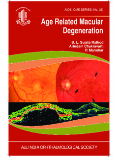
1 QU APORTA EL ESTUDIO DE LAS C lulas ganglionares EN EL glaucoma ? ANTONI DOU / JAUME RIGO / MARTA CASTANY DEPARTAMENTO glaucoma / S. OFTALMOLOG A HOSPITAL UNIVERSITARIO VALLE HEBRON glaucoma . neuropat a del nervio ptico multifactorial y adquirida, caracterizada por la p rdida de c lulas ganglionares y sus axones de la retina . El da o CFNR se sigue de cambios caracter sticos en la morfolog a del disco ptico y defectos t picos en el campo visual. - Quigley HA, Dunkelberger GR, Green WR. Retinal ganglion cell atrophy correlated with automated perimetry in human eyes with glaucoma . Am J Ophthalmol 1989;107:453-64. - Quigley HA, Miller NR, George T. Clinical evaluation of nerve fiber layer atrophy as an indicator of glaucomatous optic nerve damage. Arch Ophthalmol 1980;98:1564-71. - Sommer A, Miller NR, Pollack I, et al. The nerve fiber layer in the diagnosis of glaucoma .
2 Arch Ophthalmol 1977;95:2149 - Sommer A, Quigley HA, Robin AL, et al. Evaluation of nerve fiber layer asessment. Arch Ophthalmol 1984;102:1766-71.. C lulas ganglionares RETINIANAS (RGC) 30-50% RGC en la m cula: Densidad foveal menos variable que retina perif rica RNFL + RGC + IPL = 30-35% grosor retina OCT (RGC) MACULAR Ventaja: hay menos variabilidad de espesor GCL que en RNFL peripapilar 1 1. Grewal et al. Diagnosis of glaucoma and detection of glaucoma progression using SD optical coherence tomography. Curr Opin Ophthalm, 2013. RNFL peripapilar condicionado por: GCC: NFL + GCL + IPL tama o y forma papila vasos sangu neos atrofia peripapilar Ganglion Cell Complex FD-OCT RTVue HD-OCT Cirrus RELACI N ESTRUCTURAL-FUNCIONAL P rdida significativa de RGC puede preceder al d ficit funcional hasta en 5 a os. Sommer A, Katz J, Quigley HA, et al.
3 Clinically detectable nerve fiber atrophy precedes the onset of glaucomatous field loss. Arch Ophthalmol 1991;109:77-83. Quigley HA, Katz J, Derick RJ, Gilbert D, Sommer A. An evaluation of optic disc and nerve fiber layer examinations in monitoring progression of early glaucoma damage. Ophthalmology. 1992;99;19-28. Harwerth RS, Carter-Dawson L, Shen F, et al. Ganglion cell losses underlying visual field defects from experimental glaucoma . Invest Ophthalmol Vis Sci 1999;40:2242-50. Necesaria p rdida 17% del grosor RNFL para detectar d ficit perim trico. Wollstein. Retinal nerve fiber layer and visual function loss in glaucoma . Br J Ophthalmol, 2011 Necesaria p rdida 28% del grosor RGC para detectar d ficit perim trico. Medeiros FA, Zangwill LM, Bowd C, et al. Evaluation of retinal nerve fiber layer, optic nerve head, and macular thickness measurements for glaucoma detection usin optical coherence tomography.
4 Am J Ophthalmol 2005;139:44-55. ESTUDIO de RGC en glaucoma 1998: Primera descripci n de disminuci n en el grosor macular en glaucoma con Retinal Thickness Analyzer. Zeimer R, Asrani S, Zou s, et al. Quantitative detection of glaucomatous damage at the posterior pole by retinal thickness mapping: a pilot study. Ophthalmology 1998;105:224-31. 2003: OCT macular, confirma el hallazgo pero al compararlas, RNFL es una medida m s precisa y con mejor relaci n topogr fica del da o glaucomatoso. Lederer DE, Schuman JS, Hertzmark E, et al. Analysis of macular volumen in normal and glaucomatous eyes using Optical Coherence Tomography. Am J Ophthalmol 2003;135:838-43. Wollstein G, Schuman JS, Price LL, et al. Optical Coherence Tomography (OCT) macular and peripapillary retinal nerve fiber layer measurements and automated visual fields. Am J Ophthalmol 2004;138:218-25.
5 Bagga H, Greenfield DS, Knighton RW. Macular symmetry testing for glaucoma detection. J glaucoma 2005;14:358-63. ESTUDIO de RGC en glaucoma 2005: Capacidad diagn stica de capas internas comparable a RNFL circumpapilar. Capas internas superior a Grosor Macular en discriminaci n entre ojos sanos y glaucomatosos (p<0 049). Ishikawa H. Stein DM, Wolstein G, et al. Macular segmentation with optical coherence tomography. Invest Ophthalmol Vis Sci 2005;46:2012-17. 2009: Medida de capa de c lulas ganglionares por segmentaci n manual. Correspondencia con hallazgos funcionales. Wang M, Hood DC, Cho JS, et al. Measurement of local retinal ganglion cell layer thickness in patients with glaucoma using frequency-domain optical coherence tomography. Arch Ophthalmmol 2009;127:875-81. 2012: nuevos software de segmentaci n m s precisos FD-OCT RTVue GCC: RNFL + RGC + IPL HD-OCT Cirrus GCA: RGC-IPL HD-OCT Cirrus: protocolo GCA Medido con el protocolo de adquisici n macula 200 x 200 Cirrus HD OCT(Carl Zeiss Meditec, Dublin, CA) Base de datos normativa: 282 sujetos sanos Edad: 19-84 a os Error refractivo -12 00dp a +8 00dp Intensidad de se al >5 HD-OCT Cirrus: protocolo GCA Medido con el protocolo de adquisici n macula 200 x 200 Cirrus HD OCT(Carl Zeiss Meditec, Dublin, CA) HD-OCT Cirrus: protocolo GCA ALTA REPRODUCTIBILIDAD (Mwanza et al, 2012) HD-OCT Cirrus.
6 Protocolo GCA Capacidad diagn stica de GCA en glaucoma (Mwanza et al, 2012) GC-IPL PAR METRO ABC ROC GCIPL m nimo 0 959 GCIPL IT 0 956 GCIPL medio 0 935 GCIPL ST 0 919 GCIPL Inf 0 918 RNFL Inf 0 939 RNFL medio 0 936 RNFL Sup 0 933 C/D vertical 0 962 C/D area 0 933 ANR area 0 933 ICG Mujer (70 a) / hipotensi n arterial/ > colesterol / GNT MAVC OD 1 0 / OI 0 9 CV moderados con afectaci n paracentral Paquimetria 525/540 PIO 12-14 mmHg / 12 mmHg (latanoprost AO) ESTUDIO HOLTER (DIPS nocturnos) Pendiente de ecodoppler TSA El espesor de la retina macular total NO supera la rentabilidad diagnostica de los par metros de CFNR peri papilares An lisis GCL- IPL presenta Alta reproductibilidad Alta sensibilidad y especificidad Menor variabilidad del GCL a nivel macular que en la FNRL peripapilar Complementarios a RNFL en dx. de glaucoma En papilas an malas de morfolog a o tama o Pacientes sospechos de glaucoma defectos paracentrales focales Posible utilidad en ESTUDIO de progresi n ?
7 Mwanza et al. glaucoma diagnostic accuracy of ganglion cell-inner plexiform layer thickness: comparison with nerve fiber layer and optic nerve head. Ophthalmology. 2012. Kotowski et al. Clinical use of OCT in assessing glaucoma progression. Ophthalmic Surg Lasers, 2011. Na JH,. Detection of glaucoma progression by assessment of segmented macular thickness data obtained using spectral domain optical coherence tomography. Invest Ophthalmol Vis Sci. 2012 Jun 20;53(7):3817-26. GRACIAS POR VUESTRA ATENCI Antoni Dou Saenz de Vizmanos
