Transcription of 2 THE ANATOMY AND PHYSIOLOGY OF THE EAR …
1 2 THE ANATOMY AND PHYSIOLOGY OF THEEAR AND HEARINGP eter em. of OtolaryngologyVisiting Professor University of SingaporeUniversity of Toronto Department of OtolaryngologyToronto 5 Lower Kent Ridge RdCANADASINGAPORE INTRODUCTIONH earing is one of the major senses and like vision is important for distant warning andcommunication. It can be used to alert, to communicate pleasure and fear. It is a consciousappreciation of vibration perceived as sound. In order to do this, the appropriate signal mustreach the higher parts of the brain. The function of the ear is to convert physical vibration intoan encoded nervous impulse. It can be thought of as a biological microphone. Like amicrophone the ear is stimulated by vibration: in the microphone the vibration is transduced intoan electrical signal, in the ear into a nervous impulse which in turn is then processed by thecentral auditory pathways of the brain.
2 The mechanism to achieve this is complex. This chapterwill deal mainly with the ear, first its structure and then its function, for it is the ear that is mainlyat risk from hazardous ears are paired organs, one on each side of the head with the sense organ itself, whichis technically known as the cochlea, deeply buried within the temporal bones. Part of the ear isconcerned with conducting sound to the cochlea, the cochlea is concerned with transducingvibration. The transduction is performed by delicate hair cells which, when stimulated, initiatea nervous impulse. Because they are living, they are bathed in body fluid which provides themwith energy, nutrients and oxygen. Most sound is transmitted by a vibration of air. Vibrationis poorly transmitted at the interface between two media which differ greatly in characteristicimpedance (product of density of the medium and speed of sound within it, 'c), as for exampleair and water.
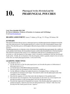
3 The ear has evolved a complex mechanism to overcome this impedancemis-match, known as the sound conducting mechanism. The sound conducting mechanism isdivided into two parts, an outer and the middle ear, an outer part which catches sound and themiddle ear which is an impedance matching device. Let us look at these parts in detail (seeFigure ). SOUND CONDUCTING The Outer EarThe outer ear transmits sound to the tympanic membrane. The pinna, that part which protrudesfrom the side of the skull, made of cartilage covered by skin, collects sound and channels it into54 ANATOMY and PHYSIOLOGY of the ear and hearingFigure The pinna and external auditory canal form the outer ear, which is separatedfrom the middle ear by the tympanic membrane. The middle ear houses three ossicles, themalleus, incus and stapes and is connected to the back of the nose by the Eustachian they form the sound conducting mechanism.
4 The inner ear consists of thecochlea which transduces vibration to a nervous impulse and the vestibular labyrinthwhich houses the organ of balance. (from Hallowell and Silverman, 1970)the ear canal . The pinna is angled so that it catches sounds that come from in front more thanthose from behind and so is already helpful in localizing sound. Because of the relative size ofthe head and the wavelength of audible sound, this effect only applies at higher frequencies. Inthe middle frequencies the head itself casts a sound shadow and in the lower frequencies phaseof arrival of a sound between the ears helps localize a sound. The ear canal is about 4centimetres long and consists of an outer and inner part. The outer portion is lined with hairyskin containing sweat glands and oily sebaceous glands which together form ear wax.
5 Hairsgrow in the outer part of the ear canal and they and the wax serve as a protective barrier and adisinfectant. Very quickly however, the skin of the ear canal becomes thin and simple and isattached firmly to the bone of the deeper ear canal , a hard cavity which absorbs little sound butdirects it to the drum head (eardrum or tympanic membrane) at its base. The outer layer of thedrumhead itself is formed of skin in continuity with that of the ear life, skin sheds and is continuously renewing. Ear canal skin grows like a fingernailfromthe depths to the exterior so that the skin is shed into the waxy secretions in the outer part55 ANATOMY and PHYSIOLOGY of the ear and hearingand falls out. This is the reason for not using cotton buds to clean the ear canal because veryfrequently they merely push the shed skin and wax deep into the canal , impacting it andobstructing hearing.
6 The ear canal has a slight bend where the outer cartilaginous part joins thebony thin skinned inner portion, so that the outer part runs somewhat backwards and the innerpart somewhat forwards. This bend is yet another part of the protective mechanism of the ear,stopping foreign objects from reaching the tympanic membrane. However it means that toinspect the tympanic membrane from the outside, one must pull the ear upwards and tympanic membrane separates the ear canal from the middle ear and is the first part of thesound transducing mechanism. Shaped somewhat like a loudspeaker cone (which is an idealshape for transmitting sound between solids and air), it is a simple membrane covered by a verythin layer of skin on the outside, a thin lining membrane of the respiratory epithelium tract on theinner surface and with a stiffening fibrous middle layer.
7 The whole membrane is less than a1/10th of millimetre thick. It covers a round opening about 1 centimetre in diameter into themiddle ear cavity. Although the tympanic membrane is often called the ear drum, technically thewhole middle ear space is the ear drum and the tympanic membrane the drum skin. The Middle Ear The middle ear is an air filled space connected to the back of the nose by a long, thin tube calledthe Eustachian tube. The middle ear space houses three little bones, the hammer, anvil andstirrup (malleus, incus and stapes) which conduct sound from the tympanic membrane to theinner ear. The outer wall of the middle ear is the tympanic membrane, the inner wall is thecochlea. The upper limit of the middle ear forms the bone beneath the middle lobe of the brainand the floor of the middle ear covers the beginning of the great vein that drains blood from thehead, the jugular bulb.
8 At the front end of the middle ear lies the opening of the Eustachian tubeand at its posterior end is a passageway to a group of air cells within the temporal bone knownas the mastoid air cells. One can think of the middle ear space shaped rather like a frying pan onits side with a handle pointing downwards and forwards (the Eustachian tube) but with a hole inthe back wall leading to a piece of spongy bone with many air cells, the mastoid air cells. Themiddle ear is an extension of the respiratory air spaces of the nose and the sinuses and is linedwith respiratory membrane, thick near the Eustachian tube and thin as it passes into the has the ability to secret mucus. The Eustachian tube is bony as it leaves the ear but as it nearsthe back end of the nose, in the nasopharynx, consists of cartilage and muscle.
9 Contracture ofmuscle actively opens the tube and allows the air pressure in the middle ear and the nose toequalize. Sound is conducted from the tympanic membrane to the inner ear by three bones, the malleus,incus and stapes. The malleus is shaped like a club; its handle is embedded in the tympanicmembrane, running from its centre upwards. The head of the club lies in a cavity of the middleear above the tympanic membrane (the attic) where it is suspended by a ligament from the bonethat forms the covering of the brain. Here the head articulates with the incus which is coneshaped, with the base of the cone articulating with the head of the malleus, also in the attic. Theincus runs backwards from the malleus and has sticking down from it a very little thin projectionknown as its long process which hangs freely in the middle ear.
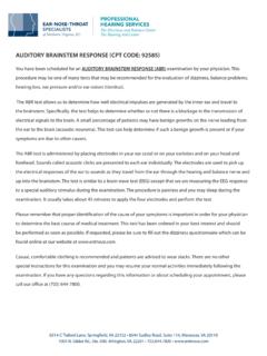
10 It has a right angle bend at itstip which is attached to the stapes(stirrup), the third bone shaped with an arch and a foot foot plate covers the oval window, an opening into the vestibule of the inner ear or cochlea,with which it articulates by the stapedio-vestibular and PHYSIOLOGY of the ear and hearingFigure The cochlea is a bony tube, filled with perilymph in which floats theendolymph filled membranous labyrinth. This separates the scala vestibuli from the scalamedia. (from Hallowell and Silverman, 1970) THE SOUND TRANSDUCING The Inner StructureThe bony cochlea is so called because it is shaped like a snail shell It has two and a half turns andhouses the organ of hearing known as the membranous labyrinth surrounded by fluid called theperilymph.