Transcription of 2100-SX, US 05M WEB SITE - Parks Med
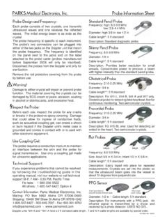
1 APPENDICESPARKS Flo-Lab operating Manual2100-SX, US 05M Appendices, Basics, Tech 10/25/06 VVVVV . 1 TECHNICAL ARTICLESBASIC operating INSTRUCTIONSUSE OF THE PENCIL PROBE IN THE DIAGNOSIS OF ARTERIAL DISEASE IN THE LIMBS1. THE PROBEThe probe consists of two crystals; one for transmitting the ultrasound waves and the other forreceiving the reflected waves. If either crystal is damaged, the probe will not work properly orwill not work at all. The crystals are covered by a material that is vulnerable to attack by ECGpaste or cream. Therefore, DO NOT use ECG paste as the contact medium between the skinand the probe. Use AQUASONIC or any gel made for ultrasonic physical therapy equipment. Inan emergency use any surgical jelly or lubricant, even petroleum jelly or mineral oil.
2 Removethe gel after use with a soft tissue. If you should find the probe with dried gel on it, wash it offunder running water. Do NOT scrape off the gel because you may damage the coating overthe crystals. Do NOT autoclave the POSITION OF THE PROBEI nvariably, people not accustomed to our probe use it incorrectly. The probe we furnish isdifferent from that of the other manufacturers and is used differently. If you hand someonethe probe and say Here, try it for yourself , they will almost always put it over their radialartery and place the probe perpendicular to the artery and perhaps with no coupling people have tried to compare our Doppler with other makes by this method. Keep inmind that you are not buying a Doppler for use on the radial artery, but for use on vesselsyou cannot feel.
3 The best testing ground is therefore in your particular area of believe our instruments will permit you to find the vessels easier, let you hear the venoussounds easier and follow the vessels better than any other device on the market, regardlessof price. But it takes some practice in order to be able to do this. We believe the arm isa good and most convenient limb for you to learn on to learn how to hold the probedepending on the depth of the artery and vein. The area about 150 mm each side of theelbow is a good place to , put some gel on the tip of the probe. The gel squeeze-bottle must be shaken downwardand then gently squeezed to get the gel to come out. Pile up about 7 mm of gel on theprobe, making certain there are no large air bubbles in the pile, because ultrasound doesnot go readily through air.
4 It needs a continuous conducting medium, and the gel is the VOLUME control fully down (counter clockwise) and turn the instrument turn up the volume. You should hear a rumbling sound if you are holding theprobe. This is caused by the vibration of the gel due to tremor in your arm. Now place theprobe over an artery in the arm about half way between the elbow and the wrist. Tilt theback of the probe toward the hand at an angle of about 45 degrees, making certain there isgel in the pathway between the probe and the skin. Move the probe and the skin sidewaysto try to find the center of the artery and the hissing noise at heart rate, which is the Dopplersound for an artery. If the sounds you hear are more or less continuous, that is simply thebackground noise of the instrument and it means that you are not over the artery.
5 Themain energy of the beam is only about as wide as the crystals in the probe, so there isn tmuch room for error in aiming the probe. For this reason you must always search the areaof the artery and tilt the probe for best Doppler Medical Electronics, Inc. Aloha, Oregon . 2 TECHNICAL ARTICLESBASIC operating INSTRUCTIONSWhen you are looking for deep arteries, or for small or obstructed arteries, you will have to turnthe VOLUME control near maximum. This also means that every time you move the head ofthe probe you are going to get some pretty big thumping noises in the earphones. Thereforeyou want to avoid moving the head of the probe with respect to the skin as much as is why you place the probe over the area where you think the artery is and then you searchfor the exact point by moving the skin with the probe and changing the angle of the probe withrespect to the skin.
6 You might wonder why these big transient noises can t be filtered. We dolimit their intensity, but we do not filter. The reason is that in the search for low-velocity bloodflow, such as in occluded arteries and in the veins, the pitch of the Doppler sounds associatedwith the blood flow are very low. Any filtering to eliminate or minimize the sounds accompanyingmovement of the probe would also reduce the response to low-velocity blood flow sounds, andof course this is DIAGNOSIS OF ARTERIAL DISEASEThe Doppler method of diagnosing arterial disease of the limbs is only one of several goodmethods. It is probably the most convenient and least expensive of the better methods. Itis only qualitative but can be made semi-quantitative by permitting you to make systolicblood pressure measurements along the leg with the aid of a proper cuff and great sensitivity of the transcutaneous Doppler can cause a doctor or technician toconclude improperly that an arterial pathway is open when it isn t.
7 Collateral flow aroundan obstruction can be well-developed, especially in the thigh, and cause pulsatile blood toflow in the distal arteries. Or a major artery may be narrowed, causing pulsatile flow mistakes in diagnosis can be avoided almost entirely by simple means and a littlebit of experience. An experienced user of the Doppler can recognize the characteristicsounds of open and obstructed arteries. Remember that Doppler sounds vary in pitch(frequency) with the velocity of blood flow. When you hear the Doppler sound on a normalartery and compare it with a normal arterial pulse-pressure wave, you will recognize thesound of the dicrotic notch, the very fast rise time of the wave and perhaps a third soundjust before the onset of a new pulse wave.
8 While the origin of these second and third wavesin the descending branch of a pulse wave may be in dispute, their absence in vessels distalto an obstruction is not disputed. So a diagnostic rule is that whenever you hear the secondand perhaps third sounds of a pulse wave of a major artery, you can be sure the artery isopen proximal to the probe. Plethysmographic studies also show a delayed crest to thewave, associated with a slower rise time to the wave when there is an obstruction the Doppler is permitting you to hear velocity changes rather than true volumechanges, the correlation is good enough to be quite valuable the opposite is not necessarily true that when you can t hear second and perhapsthird sounds the artery is obstructed proximally to the probe.
9 In the digits and smallervessels the pulse wave is smoothed out more, especially when there is somevasoconstriction. Now of course there are cases that are in doubt. If you cannot clearlyhear the second and third sounds (the third sound is frequently missing), compare with thesame artery on the other limb. If you find a radical difference in the sound of the Doppler,both in pitch and in amplitude, you are justified in being quite suspicious of the patency ofthe artery of the first limb you studied provided you are now fairly skilled at optimizing Flo-Lab operating Manual2100-SX, US 05M Appendices, Basics, Tech 10/25/06 VVVVV . 3 TECHNICAL ARTICLESBASIC operating INSTRUCTIONSA nother thing you listen for is the relative clarity of the arterial wave.
10 How well it standsout from the background noise of the instrument and perhaps the venous flow adjacent tothe artery. Move the probe a little to each side of the artery to make this estimation. In anormal person you will find that you can make the arterial pulse wave almost completelyseparate from the venous sounds by positioning of the way you really come to a final conclusion that the artery is obstructed proximal to theprobe is by measuring the systolic pressure at the ankle with an ordinary arm cuff. If youwant to measure pressure at other places on the leg you will need a special cuff, the bladderof which encircles the limb. We sell such cuffs. The method is as follows:Wrap the cuff around the ankle or slightly above it so you can get the probe on theposterior tibial and hear the arterial sounds adequately.