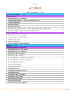
Transcription of Abdomen Sonography Examination Content Outline
1 Version: Page | 1 Abdomen Sonography Examination Content Outline ( Outline Summary) (Detailed Outline ) 1. Anatomy, Perfusion, and Function 30% Knowledge and/or skill related to anatomy, perfusion, and function Assess physical characteristics of anatomic structures (normal anatomy, anatomic variants, congenital anomalies) Biliary system Knowledge of normal anatomy, anatomic regions, and anatomic variants Knowledge of sonographic appearance of anatomic structures Ability to recognize and utilize anatomic landmarks in obtaining and documenting diagnostic images Breast Chest Liver Neck (including: thyroid, parathyroid, salivary glands, lymph nodes) Pancreas # Domain Subdomain Percentage 1 Anatomy, Perfusion, and Function Assess physical characteristics of anatomic structures Assess perfusion and function of anatomic structures 30% 2 Pathology, Vascular Abnormalities, Trauma, and Postoperative Anatomy Assess anatomic structures for pathology Assess anatomic structures for vascular abnormalities Assess anatomic structures for trauma-related abnormalities Assess aspects related to postoperative anatomy 42% 3 Abdominal Physics Apply concepts of equipment/image optimization Apply concepts of imaging artifacts 8% 4 Clinical Care, Practice, and Quality Assurance Incorporate clinical data with performed study Incorporate clinical standard/guidelines with performed study Obtain accurate measurements Assist/support during procedures 20% Version.
2 Page | 2 Penis Ability to recognize and apply proper scan technique in obtaining and documenting diagnostic images Ability to recognize, evaluate and document congenital anomalies Peritoneal cavity (including: stomach, bowel, appendix) Prostate Retroperitoneum (including: great vessels & branches) Scrotum Spleen Superficial structures (for example: abdominal wall & subcutaneous tissue) Urinary system Assess perfusion and function of anatomic structures Biliary system Knowledge of normal vascular anatomy and hemodynamics Ability to recognize appearance of normal vascular flow patterns Ability to recognize and utilize anatomic landmarks in evaluating and documenting perfusion and function Ability to recognize and apply proper scan technique in evaluating and documenting perfusion and function Chest Liver Neck (including: thyroid, parathyroid, salivary glands, lymph nodes) Penis Peritoneal cavity (including: stomach, bowel, appendix) Prostate Retroperitoneum (including: great vessels & branches) Scrotum Spleen Superficial structures (for example: abdominal wall & subcutaneous tissue) Urinary system 2.
3 Pathology, Vascular Abnormalities, Trauma, and Postoperative Anatomy 42% Knowledge and/or skill related to pathology, vascular abnormalities, trauma, and postoperative anatomy Assess anatomic structures for pathology Abdominal wall for hernia (for example: ventral, inguinal, incisional) Knowledge of etiology/pathophysiology of abnormal perfusion and function Ability to recognize ultrasound findings related to abnormalities of anatomy, perfusion, and function in obtaining and documenting diagnostic images Ability to recognize and apply proper scan technique in evaluating and documenting pathology Ability to recognize foreign bodies, infection, fluid, masses, etc. Adrenal glands for masses, hemorrhage, etc. Biliary system for infection, masses, metastatic disease, obstructions, etc. Breast for infection, abscess, masses, etc. Chest for fluid, masses, etc. Version: Page | 3 Gastrointestinal system for masses, obstruction, pyloric stenosis, intussusception, etc.
4 Knowledge of hernia types and their sonographic appearance Joints for abnormalities (for example: fluid) Liver for hepatitis, fatty infiltration, cirrhosis, neoplasm, abscess, cyst, etc. Neck (including: thyroid, parathyroid, salivary glands, lymph nodes) for diffuse parenchymal disease, inflammation, masses, etc. Pancreas for infection, masses, obstruction, etc. Penis for abnormalities Peritoneal cavity (including: stomach, bowel, appendix) for fluid Popliteal fossa for abnormalities (for example: masses, fluid) Prostate for parenchymal disease or masses (for example: benign prostatic hypertrophy) Retroperitoneum (including: great vessels & branches) for fibrosis, lymphadenopathy, etc. Scrotum for fluid, hernia, masses, infection, parenchymal disease, etc. Spleen for splenomegaly, parenchymal changes, masses, etc. Superficial structures (for example: abdominal wall, subcutaneous tissue) for foreign bodies, infection, fluid, masses, etc.
5 Urinary system for masses, obstruction, parenchymal disease, infection, etc. Assess anatomic structures for vascular abnormalities Liver for Budd-Chiari syndrome, arteriovenous fistula, portal vein thrombosis, collateralization, etc. Knowledge of anatomic and vascular changes associated with vascular abnormities Knowledge of sonographic findings associated with vascular abnormalities Ability to recognize and apply proper scan technique in evaluating and documenting vascular abnormalities Retroperitoneum (including: great vessels and branches) for aneurysm, dissection, thrombus, etc. Scrotum for torsion, varicocele, etc. Spleen for infarction, hemangiomas, etc. Version: Page | 4 Urinary system for renal artery stenosis, arteriovenous fistulas, etc. Assess anatomic structures for trauma-related abnormalities Hepatic system Knowledge of sonographic appearance as a result of trauma Ability to rapidly prioritize and evaluate sonographic findings due to trauma Ability to perform focused assessment for free fluid following a traumatic event Ability to recognize and apply proper scan technique in evaluating and documenting trauma Penis Scrotum Spleen Superficial structures (for example.)
6 Abdominal wall, subcutaneous tissue) Urinary system Focused assessment for free fluid related to traumatic events Assess aspects related to postoperative anatomy Anatomy of transplanted organs Knowledge of hemodynamics of transplanted organs Knowledge of common causes of transplant failure Ability to recognize signs of rejection Ability to adjust scan technique based on patient condition and surgical history Ability to distinguish characteristics of common anastomosis sites Ability to recognize fluid collections Ability to interpret and integrate surgical history with sonographic findings Knowledge of surgical procedures used in organ transplant Knowledge of surgical zones of the neck Ability to evaluate and document findings within surgical zones of the neck Knowledge of patterns and sonographic appearance of disease recurrence Ability to evaluate transjugular intrahepatic portosystemic shunts (TIPS) Ability to recognize and apply proper scan technique in evaluating and documenting postsurgical findings Perfusion and function of transplanted organs Complications related to organ transplants Abnormalities in postsurgical anatomy Abnormalities in postsurgical breast Abnormalities (for example: recurrent disease, lymphadenopathy) in postsurgical neck Implanted medical devices (for example: transjugular intrahepatic portosystemic shunt [TIPS]) 3.
7 Abdominal Physics 8% Knowledge and/or skill related abdominal physics Apply concepts of equipment/image optimization Use appropriate transducer (for example: curvilinear, linear, phased array) Ability to select the appropriate transducer and machine presets based on body habitus Ability to use acoustic windows creatively to optimize visualization Ability to adjust machine settings to maximize penetration while minimizing resolution loss Use two-dimensional, real-time, gray-scale imaging (for example: B-mode, compound, harmonic) Version: Page | 5 Use Doppler (for example: color, power, pulsed wave) Knowledge of appropriate application of Doppler techniques Ability to manipulate color, power, and pulsed wave settings to accurately display and measure blood flow Apply concepts of imaging artifacts Assess artifacts of gray-scale imaging (for example: shadowing, resonance, comet tail) Ability to recognize artifacts and correlate them with anatomy and pathology Ability to manipulate machine settings to enhance or minimize artifacts Assess artifacts of Doppler imaging (for example: twinkle, spectral broadening) 4.
8 Clinical Care, Practice, and Quality Assurance 20% Knowledge and/or skill related to clinical care, practice, and quality assurance Incorporate clinical data with performed study Assess indications for Examination requested Knowledge of appropriate indications and contraindications for a specific exam and/or procedure Knowledge of potential effects of patient medications on an exam or procedure Knowledge of lab values relevant to specific examinations Ability to obtain and evaluate patient history relevant to the exam Ability to assimilate patient s signs and symptoms and modify the exam/or describe the findings Ability to modify the exam based on information from other modalities Ability to localize pathology for sonographic correlation Ability to modify the exam based on real-time findings Knowledge of modalities associated with the exam being performed Ability to utilize resources, such as physicians, literature, or peers Assess relevant clinical lab values for Examination being performed Assess relevant family history and patient signs/symptoms for Examination being performed Correlate ultrasound findings with previous imaging results Evaluate images from other imaging modalities (for example.)
9 Computed tomography, magnetic resonance imaging, nuclear medicine, x-ray) Incorporate clinical standard/guidelines with performed study Communicate effectively with the patient, physician, and others, including communication of findings that require immediate action Ability to communicate with patient in a professional and appropriate manner to effectively explain procedures, deal with inappropriate behavior, and engage patient cooperation Ability to communicate using appropriate medical terminology Ability to modify exam preparation, patient position, and/or image acquisition based on patient condition and/or sonographic findings Inform patient or referring practitioner of Examination preparations (for example: fasting for biliary imaging) Maintain and protect patient confidentiality/privacy Version: Page | 6 Modify the Examination based on patient condition and/or sonographic findings Ability to recognize findings and/or situations that require immediate action and respond effectively Knowledge of appropriate patient preparation for an exam and knowledge of factors that may affect patient preparation (for example.
10 Patient history, patient condition, sequencing requirements of multiple modality exams) Knowledge of sonographer scope of practice and regulations regarding patient information and interactions Use multiple patient positions and scan planes to evaluate anatomic structures Obtain accurate measurements Obtain measurements of anatomic structures Knowledge of normal measurement ranges Knowledge of proper techniques for measuring anatomic structures Knowledge of hemodynamics Knowledge of normal and abnormal Doppler waveforms Ability to analyze Doppler measurements Ability to distinguish artifacts from actual blood flow Ability to apply knowledge of measurement techniques (for example: Doppler and gray-scale) Obtain measurements of Doppler waveforms Assist/support during procedures Obtain consent form and patient lab results prior to the procedure Knowledge of sonographer's role in obtaining consent Ability to verify and document patient consent Ability to verify correct patient, side (laterality)