Transcription of Adult Cardiac Life Support (ACLS) Recertification ...
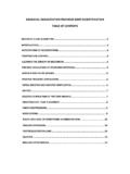
1 Adult Cardiac life Support ( acls ). Recertification Preparatory Materials 1. Adult Cardiac life Support ( acls ) Recertification . TABLE OF CONTENTS. PRIMARY AND SECONDARY ABCDs I. Primary ABCD's 03. II. Secondary ABCD's 04. AIRWAY SKILLS. I. Basic 04. II. Advanced 05. ARRHYTHMIAS 06. ELECTRICAL THERAPY 07. VASCULAR ACCESS 07. acls DRUGS 08. acls CORE CASES. I. Respiratory Arrest Case 09. II. VF Treated with CPR and AED Case 09. III. Pulseless Arrest: VF / Pulseless VT Case 10. IV. Pulseless Arrest: Pulseless Electrical Activity (PEA) Case 11. V. Pulseless Arrest: Aystole Case 11. VI. Acute Coronary Symptoms (ACS) 13. VII. Bradycardia Case 14. VIII. Unstable Tachycardia 15. IX. Stable Tachycardia 15. X. Acute Stroke Case 17. 2. What is acls ? acls is an assess then manage approach for those at risk of or in Cardiac arrest.
2 Primary and Secondary ABCD's This is a methodical assess-then-manage approach used to treat adults in respiratory distress and failure, stable and unstable arrhythmias and pulseless arrest. Algorithms are menus that guide you through recommended treatment interventions. Know the following ABCD's approach because it begins all acls case scenarios. The information you gather during the assessment will determine which algorithm you choose for the patient's treatment. I. Primary ABCDs: these refer to CPR and the AED. A. A ssess: Tap and ask: Are you OK? . A. send someone to call 911 and bring an AED. B. if alone, call 911, get an AED and return to victim Airway: Open with the head-tilt/chin lift. B. B reathing: Assess for adequate breathing. 1. if inadequate: give 2 breaths over 1 second each 2. each breath should cause a visible chest rise 3.
3 Use mouth-to-mask or barrier, bag mask - ventilation (BMV) or mouth to mouth 4. give oxygen (O2) as soon as it is available C. C irculation: Check carotid pulse for no more than 10 seconds 1. if not definitely felt, give 30 compressions in center of chest between the nipples 2. compress the chest wall 1 - 2 inches 3. one cycle of CPR is 30 compressions and 2 breaths 4. give 5 cycles of CPR (about 2 minutes). 5. minimize interruptions to compressions 6. 2 rescuers: the compressor PAUSES while 2 breaths are given 7. change compressors after 5 cycles to avoid fatigue and ineffective compressions D. D efibrillation: When an AED arrives, immediately power it on! 1. follow the voice prompts 2. use Adult pads on adults 3. II. Secondary ABCDs A. A irway: Use bag mask connected to 100% O2. 1. give each breath over 1 second each 2.
4 Compressor pauses to allow the 2 breaths to go in 3. consider inserting an advanced airway, (see Advanced Airway on page 5). B. B reathing: Look for visible chest rise during each breath 1. confirm advanced airway tube placement (see Advanced Airway on page 5). 2. secure the airway tube. 3. compressor now gives 100 continuous compressions per minute 4. ventilator gives 8-10 breaths per minute (one every 6-9 seconds). C. C irculation: Obtain vascular access with an IV(intravascular) or 10 (intraosseous) cannula 1) give drugs as recommended per algorithm. D. D iagnosis: Why is the patient in the rhythm? Look for any possible causes to treat: 6 Hs 5 Ts H ypoxia T amponade H ypovolemia T ension pneumothorax H ypothermia T oxins poisons, drugs H ypoglycemia T hrombosis coronary (AMI) pulmonary (PE). H ypo / H yperkalemia T rauma H ydrogen ion (acidosis).
5 Airway Skills I. Basic Airway A. Oxygen B. Open the Airway 1. use the head - tilt chin lift when assessing for adequate breathing 2. use a jaw thrust for unresponsive unwitnessed, trauma or drowning victims if unable to open the airway with a jaw thrust, use head-tilt chin lift C. Maintain 1. insert an oropharyngeal airway when unconscious with no cough or gag reflex 2. insert a nasopharyngeal airway when a cough or gag reflex is present (better tolerated). D. Ventilate: give each breath over 1 second using enough volume to see the chest rise 1. 2-rescuer CPR: give 2 breaths during the pause following 30 compressions 2. Rescue breathing: when a pulse is present, give 10-12 breaths/minute (one each 5-6. seconds). 4. II. Advanced Airway A. Laryngeal Mask Airway (LMA): requires the least training for insertion 1.
6 Inserts blindly into the hypopharynx 2. regurgitation and aspiration are reduced but not prevented 3. confirm placement: see chest rise and listen for breath sounds over lung fields 4. contraindications: gastric reflux, full stomach, pregnancy or morbid obesity B. Combitube: requires more training for insertion than the LMA. 1. inserts blindly into esophagus (80% of the time) or the trachea 2. ventilation can occur whether the tube is the esophagus or the trachea 3. confirm placement: clinical exam and a confirmation device (see devices). 4. contraindications: gag reflex, esophageal disease, caustic ingestion, under 16 years of age or under 60 inches in height C. Endotracheal Tube (ETT): requires the most training, skill and frequent retraining for insertion 1. inserts by direct visualization of vocal chords 2.
7 Isolates the trachea, greatly reduces risk of aspiration and provides reliable ventilation 3. high risk of tube displacement or obstruction whenever patient is moved 4. confirm placement, clinical exam and a confirmation device (see devices). D. Confirm tube placement 1. Clinical Assessment a. look for bilateral chest rise b. listen for breath sounds over stomach and the 4 lung fields (left and right anterior and midaxillary). c. look for water vapor in the tube (if seen this is helpful but not definitive). 2. Devices a. End-Tidal CO2 Detector (ETD): if weight > 2 kg i. attaches between the ET and Ambu bag; give 6 breaths with the Ambu bag ii. litmus paper center should change color with each inhalation and each exhalation original color on inhalation = OK O2 is being exhaled: expected color change on exhalation = CO2!
8 ! Tube is in trachea iii. Trouble shooting: original color on exhalation =. litmus paper is wet replace ETD, or tube is not in trachea remove ET, or Cardiac output is low during CPR. b. Esophageal Detector (EDD): if weight > 20 kg and in a perfusing rhythm i. Resembles a turkey baster compress the bulb and attach to end of ET. bulb inflates quickly - tube is in the trachea bulb inflates poorly - tube is in the esophagus ii. No recommendation for its use in Cardiac arrest 5. Arrhythmias Pulseless Rhythms (Arrest Rhythms). Shockable Non-Shockable VF (Ventricular Fibrillation) PEA (Pulseless Electrical Activity). VT (Ventricular Tach Pulsless) Asystole (Silent Heart). Torsades de Pointes Perfusing Rhythms (Non-Arrest Rhythms). Bradycardia Tachycardias: Narrow QRS. Sinus Bradycardia Regular Rhythms: Junctional Rhythm Sinus Tachycardias Idioventricular Rhythm Atrial Flutter Supraventricular Artioventricular Block: Tachycardia 1st Degree Junctional Tachycardia 2nd Degree: Mobitz Type I Irregular Rhythms: (Wenckebach) Atrial Flutter Mobitz Type II Atrial Fibrillation rd 3 Degree Multifocal Atrial (Complete Heart Block) Tachycardia Tachycardias: Wide QRS.
9 Regular Rhythm: Ventricular Tachycardia . monomorphic Ventricular Tachycardia . polymorphic Torsades des pointes 6. Electrical Therapy I. Defibrillation: high energy shocks with manual defibrillator: ECC handbook, p. 9. A. recommended shock dose: biphasic = 120 200 J (per manufacturer). B. recommended shock dose: monophasic = 360 J. II. Synchronized Cardioversion: timed low energy shocks: ECC handbook p. 14. A. timed to QRS to reduce risk of R-on-T : a shock that hits the T wave may cause VF. III. Transcutaneous Pacer: noninvasive emergent bedside pacing: ECC handbook, p. 62. A. apply pacer pads B. verify pacer capture Vascular Access Peripheral: preferred in arrest due to easy access and no interruption in CPR. A. use a large bore IV catheter B. attempt large veins: anticubital, external jugular, cephalic, femoral C.
10 Can take 1-2 minutes for IV drugs to reach the central circulation D. follow IV drugs with a 20 ml bolus of IV fluid and elevate extremity for 10-20 seconds Intraosseous (IO): inserts into a large bone and accesses the venous plexus A. may use if unable to obtain intravascular access B. drug delivery is similar to that via a central line C. safe access for fluids, drugs, blood samples D. commercial kits are available for IO access E. drug doses are the same as when given IV. Central Line: not needed in most resuscitations A. insertion requires interruption of CPR. B. if a central line is already in place and patent, it can be used Endotracheal: some drugs may be given via the ETT in the absence of a IV/IO. A. drug delivery is unpredictable thus IV/IO delivery is preferred B. drug blood concentration stay lower than when given IV.
