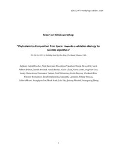
Transcription of Analysis and Isolation of Adipocytes by Flow …
1 CHAPTER FIFTEENA nalysis and Isolation ofAdipocytes by Flow CytometrySusan M. Majka*, Heidi L. Miller , Karen M. Helm{, Alistaire S. Acosta{,Christine R. Childs{, Raymond Kong}, Dwight J. Klemm ,1*Department of Medicine, Vanderbilt University, Nashville, Tennessee, USA Department of Medicine, University of Colorado Anschutz Medical Campus, Aurora, Colorado, USA{Cancer Center Flow cytometry Core, University of Colorado Anschutz Medical Campus, Aurora, Colorado,USA}Amnis Corporation, Seattle, Washington, USA1 Corresponding author: e-mail address: of Adipocytes by Collagenase Digestion of Adipose of Single Cell Suspensions from Adipose Tissue for Flow Deep Red neutral lipid staining of lipid antibody staining to exclude stromal/vascular cells Violet staining to identify events with nuclei and distinguishsinglets from and Sorting of Adipocytes by Flow XDP of Adipocytes from stromal cells based on cell size: FSC versus of single cells (singlets) from cell of events containing lipid of stromal/vascular of adipocyte Isolation and Isolation of Adipocytes via flow cytometry is particularly useful to studytheir biology.}}
2 However, the adoption of this technology has often been hamperedby the presence of stromal/vascular cells in adipocyte fractions prepared fromcollagenase-digested adipose tissue. Here, we describe a multistep staining methodand gating strategy that effectively excludes stromal contaminants. Initially, we set aMethods in Enzymology, Volume 537#2014 Elsevier 0076-6879 All rights optimized to the size and internal complexity of Adipocytes . Exclusion of cell aggre-gates is then performed based on fluorescence of a nuclear stain followed by positiveselection to collect only those cell events containing lipid droplets. Lastly, negativeselection of cells expressing stromal or vascular lineage markers removes any remainingstromal contaminants.
3 These procedures are applicable to simple Analysis of adipocytesand their subcellular constituents by flow cytometry as well as Isolation of Adipocytes byflow INTRODUCTIONFlow cytometry is a powerful technique that provides the ability torapidly measure multiple cellular parameters across single cell suspensionsand large cell populations with tremendous precision. During cytometry , Analysis of light scatter can distinguish different cells based on their size,shape, and internal complexity. The presence and amount of specific intra-cellular and cell surface molecules can be measured with antibodies orligands conjugated with fluorescent probes. Likewise, fluorescent indicatorsare available to measure the transport of ions across cellular membranes, aswell as assess mitochondrial activity and other metabolic parameters.
4 Specificcell subtypes can be isolated from mixed populations of single cell suspen-sions on specialized cytometers called sorters. In spite of the tremendousanalytical power and advantages afforded by flow cytometry , this technologyhas been underutilized in the study of adipocyte general assumption hampering the broad scale adoption of flow cyto-metry to the study of Adipocytes is that they are too large (50 200mm) andfragile to effectively analyze and sort using modern benchtop flowcytometers. However, the internal diameter of the fluidics of modern flowcytometers ranges from 150 to 250mm large enough to accommodate allbut the largest fat cells. Moreover, typical flow pressures range from only 5 to10 psi and the laminar flow within the cytometer exerts minimal shear stresson cells.
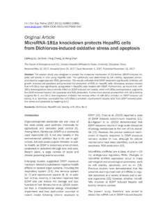
5 In addition, Adipocytes are fairly deformable. Their shape is largelydefined by large cytoplasmic lipid droplets, which contain liquid triglycerideat room or higher temperature, rather than cytoskeletal elements. In collab-oration with Amnis Corporation (Seattle, WA) we have acquired brightfieldimages of intact unilocular and multilocular Adipocytes (and cell debris) inthe fluidics stream of an ImageStream X Imaging Flow Cytometer(Fig. ).282 Susan M. Majka et Adipocytes and collecting them for study via flow sorting is a bitmore complex than Analysis alone. Sorting exposes cells to higher flow pres-sures, smaller fluidic diameters, and therefore, increased shear stress. Withinthe sorter, cells are encapsulated within droplets of sheath fluid.
6 The trajec-tory of the droplets is controlled by imparting them with an electric path of the charged droplets is deflected toward an oppositely chargedplate so that individual droplets may be captured in separate tubes. However,the high flow pressures and relatively small tip diameters (70 100mm)required to produce the droplets may result in shear-induced adipocyte spite of these conditions we find that a small percentage of fat cells survivesorting as determined by microscopy of the sorted cells (Fig. ). More-over, lysis of Adipocytes during sorting can actually facilitate recovery ofFigure images of unilocular and multilocular Adipocytes , and cell debrisacquired with an ImageStream X Imaging Flow Cytometer at Amnis Corporation.
7 Theimages demonstrate the feasibility of using flow cytometry to study intact Cytometric Analysis of Adipocytesintact nuclei (Majka et al., 2010), nucleic acids, and other cellular constitu-ents for further Analysis (Majka et al., 2010; Schaedlich, Knelangen, Santos,Fischer, & Santos, 2010).Applying these concepts, we and other laboratories have begun toexploit flow cytometry to study aspects of adipocyte biology with consider-able success. For example,Shi and Kandror (2008)andBruzzone et al.(2012)developed flow cytometry techniques to measure the degree of trans-location of the insulin-stimulated glucose transporter, Glut4, to the mem-brane of 3T3-L1 et al. (2012)measured mitochondrialmembrane potential and production of reactive oxygen species in Lyrm1-depleted 3T3-L1 Adipocytes , whileFesty et al.
8 (2005)used flow cytometryto assess surface protein expression between human primary Adipocytes andstromal cells. Other laboratories have employed flow cytometry to measurelipid accumulation in Adipocytes (Lee, Chen, Wiesner, & Huang, 2004),assess production of Toll-like receptor 2 and tumor necrosis factorainprimary Adipocytes (Murakami, Bujo, Unoki, & Saito, 2007), and trackthe differentiation of rat primary preadipocytes in response to anandamide(Karaliota, Siafaka-Kapadai, Gontinou, Psarra, & Mavri-Vavayanni, 2009).We exploited flow cytometry and fluorescence deconvolution microscopyto demonstrate the production of Adipocytes from bone marrow-derivedprogenitor cells (Crossno, Majka, Grazia, Gill, & Klemm, 2006; Majkaet al.)
9 , 2010, 2012). Additionally, we found that flow sorting of primarymouse Adipocytes was a highly efficient means to isolate intact nuclei forcytogenetic Analysis (Majka et al., 2010) and RNA for global gene et al. (2010)used similar methods to recover RNAfrom embryonic stem cell-derived Adipocytes for RT-PCR images of an intact unilocular and an intact multilocular adipo-cyte after flow sorting. Free-floating Adipocytes were sorted on a MoFlo XDP instrumentequipped with a 70-mm tip. Adipocytes were collected in a 12 75-mm polypropylenetube containing 150ml of flow buffer. Images were acquired with a conventional M. Majka et obesity and metabolic diseases linked to adipose tissue continue toplace health and financial burdens on society, scientists have begun to addresscomplicated questions about adipocyte production and lineage, fat cell turn-over, apoptosis and senescence.
10 Flow cytometry is an important addition tothe arsenal of sophisticated techniques that are now employed in these stud-ies. The ability of flow cytometry to screen multiple adipocyte parametersover large cell populations is an advantage over microscopic , even the best commercial laser confocal systems are limited toa nominal resolution of approximately 200 nm, making it difficult to discernthe orientation and relationship between structures and markers in tissuesections and whole mounts. Flow cytometry overcomes this limitation byfacilitating Analysis of individual cells or events. Imaging flow cytometers,including the ImageStream X (Amnis, Seattle, WA) combine the large pop-ulation/multiple parameter advantages of traditional flow cytometry , withthe ability to acquire brightfield images of Adipocytes in the flow , modern flow cytometers may exceed the ability of laser confocalmicroscopes to detect diffuse fluorescent signals ( , cytosolic EGFP), sincefluorescence is integrated over the entire cell or event instead of final remaining challenge to using flow cytometry in the study of adi-pocytes is the presence of contaminating stromal/vascular cells.