Transcription of Anatomy and Physiology of Domestic Animals
1 Anatomy and Physiology of Domestic Animals6 Bones and skeletal systemContentsBones Introduction Classifi cation of BonesBone Structure Gross Anatomy Microscopic Anatomy of Bone Chemical Composition of Bone Hematopoietic Tissue in BoneBone Development Intramembranous Ossifi cation Endochondral Ossifi cationBone Growth, Remodeling, and Repair Bone Growth Bone Remodeling and Repair Repair of FracturesHomeostatic Imbalances of Bone Osteomalacia and Rickets Parturient Paresis (Milk Fever) Egg-Laying Fatigue in BirdsBones and Skeleton Markings on Bones Skeleton Skeletal Cartilage Skeleton Classifi cationAxial Skeleton The Skull The Vertebral ColumnAppendicular Skeleton Thoracic Limb Pelvic LimbAvian SkeletonJoints Types of Joints Synovial JointsSpecifi c Joints Intervertebral Articulations Thoracic Limb Pelvis Pelvic LimbBonesIntroductionOsteology is the study of bones.
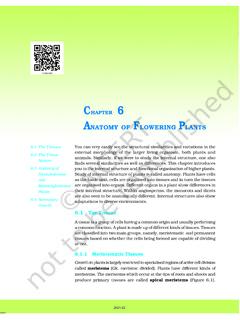
2 The skeleton pro-vides the basic scaffolding for the body. The skeletal system includes the bones, and the cartilage, liga-ments, and connective tissues that hold everything cation of bonesThe human skeleton contains 206 major bones whereas the number of bones in different Animals varies. The bones can be classifi ed into fi ve categories including long bones, short bones, fl at bones, irregular bones, and sesamoid bones (Fig. ). Long bones. These are bones that are longer than they are wide. Some of the bones of the limbs are 133134 Anatomy and Physiology of Domestic animalslong bones. Long bones are characterized by an elongated shaft and somewhat enlarged extremi-ties that bear articular surfaces. Examples of long bones include the humerus, radius, femur, tibia, metacarpals, and metatarsals.
3 Short bones. Short bones are generally cube-shaped, and examples include the carpal and tarsal bones. Flat bones. Flat bones, as the name implies, are thin and fl attened. They include two plates of compact bone separated by cancellous or spongy bone. Examples include the sternum, ribs, scapula, and certain skull bones. Irregular bones. These are complex and irregu-larly shaped bones. Examples include the verte-brae and certain facial bones. Sesamoid bones. Sesamoid bones are small bones embedded in a tendon and resemble the shape of a sesame seed. Examples include the patella, and proximal and distal sesamoid bones of the structureGross anatomyEach bone consists of compact bone and cancellous bone. Compact bone, also called dense or cortical bone, is a term describing solid-looking bone.
4 Compact bone is found on the surface of bones forming a pro-tective outer coating; cancellous bone is found on the bone, also called spongy bone, consists of a network of pieces of bone called trabeculae or spicules, interspersed with spaces fi lled with red or yellow bone marrow. Spongy bone predominates in short, fl at, and irregular bones, as well as in the epiph-yses of long bones. It is also found as a narrow lining of the medullary cavity of the diaphysis of long bones. The epiphyses consist mostly of cancellous bone with a thin outer coat of compact developing long bones, the shaft is called the diaphysis and each extremity is called an epiphysis (pl. = epiphyses) (Fig. ). The epiphysis consists mostly of cancellous bone with a thin outer coat of compact bone. It is generally enlarged relative to the diaphysis.
5 The metaphysis is the joining point of the diaphysis and epiphysis. Between the diaphysis and epiphysis of growing bones is a fl at plate of hyaline cartilage called the epiphyseal plate. After growth is complete, the plate is replaced by the epiphyseal line. The medullary cavity (medulla, innermost part ) is the space in the diaphysis containing bone marrow. At the joint surface on the bone is an articular surface consisting of a smooth layer of hyaline cartilage that covers the epiphysis where one bone forms a joint with another fi brous covering surrounding that part of the bone not covered with articular cartilage is called the periosteum. It consists of dense irregular connective tissue. Its innermost layer consists of an osteogenic layer containing osteoblasts (bone germinators) that make new bone, and osteoclasts that break down bone.
6 The periosteum contains nerve fi bers, lymphatic vessels, and blood vessels that supply the bone. The periosteum is attached to the underlying bone by Sharpey s fi bers extending from the fi brous layer into the bone matrix. There is a high density of Sharpey s fi bers where tendons and ligaments attach to the internal surfaces of the bone are covered with the endosteum. The endosteum lines the medullary cavity in long bones and covers the trabeculae of spongy , irregular and fl at bones vary in the propor-tion of compact and cancellous (Fig. ). Furthermore, these bones also do not have a shaft or epiphyses. They contain bone marrow between their trabeculae, but there is no bone marrow cavity. The internal spongy layer in fl at bones is called the diplo (folded).
7 Microscopic Anatomy of boneThere are four major cell types found in bone (Fig. ). Osteocytes are the mature cells within bone that account for most of the population of bone cells. They are found within a lacuna (see next section, Compact Bone ). Osteoblasts are cells that secrete the extracel-lular matrix on bone. They secrete collagen and ground Irregular bone(vertebrae)Long bone(femur)Flat bone(rib)Short bone(carpal)Sesamoid bone(patella)Fig. Types of bones. Examples of the various types of bones as found in the pig and skeletal system 135substance that makes up unmineralized bone, called osteoid. Once these cells get embedded within the matrix, they become osteocytes. Osteoclasts are cells involved in resorption of bone, and are therefore present in areas where bone is being removed. Osteo-clasts are giant multinucleated cells.
8 Bone also con-tains a small number of mesenchymal cells known as osteoprogenitor cells. These are stem cells that can produce osteoblasts, and are therefore important in fracture repair. They are located in the inner, cellular layer of the periosteum, the endosteum that lines the marrow cavity, and the lining of vascular passage-ways in the boneAlthough compact bone appears solid to the unaided eye, microscopically it contains considerable detail. The structural unit of compact bone is the osteon, or Haversian system (Fig. ). Each osteon appears as a cylindrical unit consisting of 3 20 concentric lamellae of bone matrix surrounding the central osteonal canal (Haversian canal, or central canal) that runs parallel to the long axis of the bone. The lamellae are like paper towels wrapped around a cardboard roll ( , osteonal canal).
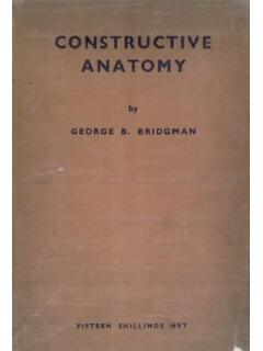
9 The osteonal canal contains the vascular and nerve supply of the osteon. The osteonal canals run parallel to the long axis of the bone, and they carry small arteries and second group of canals, called perforating or Volk-mann s, or lateral, canals, run at right angles to the long axis of the bone. These canals connect the blood vessel and nerve supply of the periosteum with that in the osteonal canal. These canals are lined with bone formation, osteoblasts secrete the bone matrix. However, osteoblasts maintain contact with Spongy boneMedullary cavityCompact boneYellow marrowPeriosteumArticularCartilageSpongy boneEpiphysisDiaphysisEpiphysisSpongy boneBACompact boneMedullary cavityMetaphysisMetaphysisEpiphysisMetap hysisDiaphysisMetaphysisEpiphysisArticul arcartilageRed MarrowFig. Anatomy of long bones.
10 A) Using the femur as an example of a long bone, the epiphysis is the enlarged area at either end of the bone while the diaphysis is the long shaft in the middle portion of the bone. The metaphysis is the joining point between the epiphysis and diaphysis. The periosteum is the fi brous covering around the outside of the bone not covered with articular cartilage. The endosteum is the fi brous and cellular tissue lining the medullary cavity of the bone. B) Cross section of an equine humerus showing exterior and interior Anatomy and Physiology of Domestic animalsone another via connections containing gap junctions. As the matrix hardens, the osteoblasts become trapped within it, thus forming the lacunae and canaliculi. The osteoblasts become osteocytes, or mature bone , the spider-shaped mature bone cells, are found in lacunae, the small cavities at the junctions of the lamellae.