Transcription of Anatomy and Physiology of the Skin - ONS
1 1 Anatomy and Physiology of the skin Paul Kolarsick, BS, Maria Ann Kolarsick, MSN, ARNP-C, and Carolyn Goodwin, APRN-BC, FNPCHAPTER 1 IntroductionThe skin is the largest organ of the body, accounting for about 15% of the total adult body weight. It performs many vital functions, including protection against external physical, chemical, and biologic assailants, as well as prevention of ex-cess water loss from the body and a role in thermoregulation. The skin is continuous, with the mucous membranes lining the body s surface (Kanitakis, 2002). The integumentary system is formed by the skin and its derivative structures (see Figure 1-1). The skin is composed of three layers: the epidermis, the dermis, and subcutaneous tissue (Kanitakis, 2002). The outermost level, the epidermis, consists of a specific constellation of cells known as keratinocytes, which function to synthesize keratin, a long, threadlike protein with a protective role.
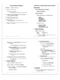
2 The middle layer, the dermis, is fundamentally made up of the fibrillar structural protein known as collagen. The der-mis lies on the subcutaneous tissue, or panniculus, which contains small lobes of fat cells known as lipocytes. The thickness of these layers varies considerably, depending on the geographic location on the Anatomy of the body. The eyelid, for example, has the thinnest layer of the epidermis, measuring less than mm, whereas the palms and soles of the feet have the thickest epidermal layer, measuring approximately mm. The dermis is thickest on the back, where it is 30 40 times as thick as the overlying epidermis (James, Berger, & Elston, 2006). Figure 1-1. Cross-Section of skin and PanniculusNote. From Andrews Diseases of the skin : Clinical Dermatology (10th ed., p. 1), by James, Berger, and Elston, 2006, Phila-delphia: Elsevier Saunders. Copyright 2006 by Elsevier Saunders.
3 Reprinted with nerve endingDermispapillaryreticularSebaceous glandArrector pili muscleHair shaftPacini nerve endingSubcutaneous tissueApocrine unitStraight ductCoiled glandEccrine sweat unitStraight ductStraight ductCoiled ductEccrine glandDermal vasculatureSuperficial plexusDeep plexusSKIN CANCER2 EpidermisThe epidermis is a stratified, squamous epithelium layer that is composed primarily of two types of cells: keratinocytes and dendritic cells. The keratinocytes differ from the clear dendritic cells by possessing intercellular bridges and ample amounts of stainable cytoplasm (Murphy, 1997). The epidermis harbors a number of other cell populations, such as melanocytes, Langerhans cells, and Merkel cells, but the keratinocyte cell type comprises the majority of the cells by far. The epidermis commonly is divided into four layers according to keratino-cyte morphology and position as they differentiate into horny cells, including the basal cell layer (stratum germinativum), the squamous cell layer (stratum spinosum), the granular cell layer (stratum granulosum), and the cornified or horny cell layer (stratum corneum) (James et al.)
4 , 2006; Murphy) (see Figure 1-2). The lower three layers that constitute the living, nucleated cells of the epidermis are sometimes referred to as the stratum malpighii and rete malpighii (Murphy).The epidermis is a continually renewing layer and gives rise to derivative structures, such as pilosebaceous apparatuses, nails, and sweat glands. The basal cells of the epidermis un-dergo proliferation cycles that provide for the renewal of the outer epidermis. The epidermis is a dynamic tissue in which cells are constantly in unsynchronized motion, as differing individual cell populations pass not only one another but also melanocytes and Langerhans cells as they move toward the surface of the skin (Chu, 2008).KeratinocytesAt least 80% of cells in the epidermis are the ectodermally derived keratinocytes. The differentiation process that occurs as the cells migrate from the basal layer to the surface of the skin results in keratinization, a process in which the kerati-nocyte first passes through a synthetic and then a degradative phase (Chu, 2008).
5 In the synthetic phase, the cell builds up a cytoplasmic supply of keratin, a fibrous intermediate filament arranged in an alpha-helical coil pattern that serves as part of the cell s cytoskeleton. Bundles of these keratin filaments converge on and terminate at the plasma membrane forming the intercellular attachment plates known as desmosomes. During the degradative phase of keratinization, cellular organelles are lost, the contents of the cell are consolidated into a mixture of filaments and amorphous cell envelopes, and the cell finally is known as a horny cell or corneocyte. The process of maturation resulting in cell death is known as terminal differentiation (James et al., 2006).Basal LayerThe basal layer, also known as the stratum germinativum, contains column-shaped keratinocytes that attach to the base-ment membrane zone with their long axis perpendicular to the dermis. These basal cells form a single layer and adhere to one another as well as to more superficial squamous cells through desmosomal junctions (Murphy, 1997).
6 Other dis-tinguishing features of the basal cells are their dark-staining oval or elongated nuclei and the presence of melanin pigment transferred from adjoining melanocytes (Murphy).The basal layer is the primary location of mitotically active cells in the epidermis that give rise to cells of the outer epidermal layers. However, not all basal cells have the potential to divide (Jones, 1996; Lavker & Sun, 1982). Epidermal stem cells in the basal layer are clonogenic cells with a long lifespan that progress through the cell cycle very slowly under normal conditions. Hy-perplasiogenic conditions, such as wounding, can increase the number of cycling cells in the epidermis by stimulating division of stem cells. DNA damage caused by carcinogenic agents may mutate cell proliferation machinery and can also affect the rate of cellular division. Migration of a basal cell from the basal layer to the cornified layer in humans takes at least 14 days, and the transit through the cornified layer to the outermost epidermis requires another 14 days (Chu, 2008).
7 Squamous Cell LayerOverlying the basal cell layer is a layer of the epidermis that is 5 10 cells thick and known as the squamous cell layer Figure 1-2. Three Basic Cell Types in the EpidermisThe three basic cell types in the epidermis include keratinocytes (some labeled K) and Langerhans cells (L) in the Malpighian layer and melanocytes (M) in the basal layer. Arrows point to the basement membrane zone, which separates the basal layer of the epidermis from the underlying dermis (D).Note. From Andrews Diseases of the skin : Clinical Dermatology (10th ed., p. 4), by James, Berger, and Elston, 2006, Philadelphia: Elsevier Saunders. Copyright 2006 by El-sevier Saunders. Reprinted with 1. Anatomy AND Physiology OF THE SKIN3or stratum spinosum (Murphy, 1997). The squamous layer is composed of a variety of cells that differ in shape, structure, and subcellular properties depending on their location.
8 Supra-basal spinous cells, for example, are polyhedral in shape and have a rounded nucleus, whereas cells of the upper spinous layers are generally larger in size, become flatter as they are pushed toward the surface of the skin , and contain lamellar granules (Chu, 2008). Lamellar granules are membrane-bound organelles containing glycoproteins, glycolipids, phospholip-ids, free sterols, and a number of acid hydrolases, including lipases, proteases, acid phosphatases, and glycosidases. The abundance of hydrolytic enzymes indicates that the lamel-lar granules are a type of lysosome. Although the lamellar granules primarily are active in cells at the interface between the granular and cornified layers, they also function in cells of the upper spinous layer to deliver precursors of stratum corneum lipids into the intercellular space (Haake & Hol-lbrook, 1999).Intercellular spaces between spinous cells are bridged by abundant desmosomes that promote mechanical coupling be-tween cells of the epidermis and provide resistance to physical stresses.
9 Organized concentrically around the nucleus, keratin filaments in the cytoplasm are bound to desmosomal plaques at one end and remain free at the end closer to the nucleus (Murphy, 1997). The desmosomal plaques are composed of six polypeptides found on the cytoplasmic side of the cell membrane that are important in the regulation of the calcium required for desmosomal assembly and maintenance (Fairley, Scott, Jensen, Goldsmith, & Diaz, 1991; Hennings & Holbrook, 1983; Lin, Mascaro, Liu, Espana, & Diaz, 1997). The spine-like appearance of the numerous desmosomes along cell margins is where the stratum spinosum derives its name (Chu, 2008).Gap junctions are another type of connection between epi-dermal cells. Essentially forming an intercellular pore, these junctions allow for physiologic communication via chemi-cal signals that is vital in the regulation of cell metabolism, growth, and differentiation (Caputo & Peluchetti, 1977).
10 Granular LayerThe most superficial layer of the epidermis containing living cells, the granular layer or stratum granulosum, is composed of flattened cells holding abundant keratohyaline granules in their cytoplasm. These cells are responsible for further synthesis and modification of proteins involved in keratinization (Chu, 2008). The granular layer varies in thick-ness in proportion to that of the overlying horny cell layer. For example, under thin cornified layer areas, the granular layer may be only 1 3 cell layers in thickness, whereas under the palms of the hands and soles of the feet the granular layer may be 10 times this thickness. A very thin or absent granular layer can lead to extensive parakeratosis in which the nuclei of keratinocytes persist as the cells move into the stratum corneum, resulting in psoriasis (Murphy, 1997). The keratohyaline granules are deeply basophilic and irregular in shape and size, and they are necessary in the formation of both the interfibrillary matrix that holds keratin filaments together and the inner lining of the horny cells.