Transcription of ANATOMY OF LUNGS - College of Arts & Sciences
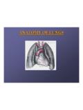
1 ANATOMY OF LUNGSANATOMY OF LUNGS --1. Gross ANATOMY of Lungs2. Surfaces and Borders of Lungs3. Hilum and Root of Lungs4. Fissures and Lobes of Lungs5. Bronchopulmonarysegments6. Histopathology of supply of of Lungs10. Nerve supply of Lungs11. Pleura12. MediastinumGROSS ANATOMY OF LUNGSGROSS ANATOMY OF LUNGSL ungs are a pair of respiratory organs situated in a thoracic cavity. Right and left lung are separated by the Young brownAdults -- mottled black due to deposition of carbon particlesWeight-Right lung - 600 gmsLeft lung - 550 gmsTHORACIC CAVITYTHORACIC CAVITYSHAPE-ConicalApex(apex pulmonis)Base(basis pulmonis)3 Borders-anterior (margo anterior)-posterior (margo posterior)- Inferior (margo inferior)2 Surfaces-costal (facies costalis)- medial (facies mediastinus)- anterior (mediastinal)- posterior (vertebral)APEXAPEXB luntBluntLies above the level of Lies above the level of anterior end of 1anterior end of 1 Reaches 1--2 cm above 2 cm above medial 1/3medial 1/3rdrdof cervical bGrooved by--SubclavianSubclavianartery artery SubclavianSubclavianveinveinBASEBASES emilunarSemilunarand on dome of on dome of sided dome is higher than sided dome is higher than BORDERANTERIOR BORDER to the anterior Corresponds to the anterior ((CostomediastinalCostomediastinal) line of pleural reflection.)
2 Line of pleural is deeply notched in the left lung posterior It is deeply notched in the left lung posterior to 5to 5ththcostal cartilage by the pericardium costal cartilage by the pericardium and extends vertically downwards to formand extends vertically downwards to formLingulaLingula. This is called . This is called cardiac cardiac notchnotch(percussion in this(percussion in thisarea gives a dull area gives a dull note as compared to dull note obtained over note as compared to dull note obtained over lung).lung).INFERIOR BORDERINFERIOR BORDERThin and sharp Thin and sharp It It seperatesseperatesthe base of lung from the the base of lung from the costal surface and extends into costal surface and extends into BORDERPOSTERIOR BORDERT hick and ill definedThick and ill definedFits into deep Fits into deep from C7 to from C7 to OF THE LUNG1. Costal Surface- It is in contact with costal pleura and overlying thoracic Medial Surface- Posterior / Vertebral Part- Anterior / Mediastinal PartRelations of Posterior Part1.
3 Vertebral Part2. Intervertebral Discs3. Posterior Intercostal Vessels4. Splanchic NervesRELATIONS OF ANTERIOR PARTRELATIONS OF ANTERIOR PARTRIGHT SIDERIGHT atriumRight part of RVSmall part of Right brachiocephalicbrachiocephalicvein(lower part)vein(lower part) Right Right phrenicphrenicnervenerveLEFT SIDELEFT ventricleLeft trunkPulmonary of AortaArch of thoracic Descending thoracic Left ductThoracic Left Left Left recurrent Left recurrent laryngeal nervelaryngeal nerveHILUMHILUMIt is a large depressed area that lies It is a large depressed area that lies near the centre of the medial surface. near the centre of the medial surface. Various structures enter and leave the Various structures enter and leave the lung via its via its OF THE LUNGROOT OF THE LUNGThe root is enclosed in The root is enclosed in a short tubular sheet of a short tubular sheet of pleura that joins the pleura that joins the pulmonary and pulmonary and mediastinalmediastinalparts of parts of pleura.
4 It extends pleura . It extends inferiorly as a narrow inferiorly as a narrow fold fold --The pulmonary The pulmonary lies opposite of the It lies opposite of the bodies of 5th, 6th and bodies of 5th, 6th and 7th thoracic vertebra7th thoracic vertebraSTRUCTURES OF THE ROOTSTRUCTURES OF THE ROOTP rincipal Bronchus on the Principal Bronchus on the left and HyparterialHyparterialon the right the right pulmonary artery .One pulmonary artery .Two pulmonary veins Two pulmonary veins --SuperiorSuperiorInferiorInferiorBronch ial arteriesBronchial arteriesOne on right sideOne on right sideTwo on left sideTwo on left sideBronchial veinsBronchial veinsAnterior and Anterior and posterior pulmonary posterior pulmonary plexus of of Bronchopulmonary LymphnodesLymphnodesAreolar OF STRUCTURES IN ARRANGEMENT OF STRUCTURES IN THE ROOTTHE ROOTBEFORE BACKWARDSBEFORE BACKWARDS1.
5 Superior pulmonary Superior pulmonary Pulmonary Pulmonary OF STRUCTURES IN ARRANGEMENT OF STRUCTURES IN THE ROOTTHE ROOTABOVE DOWNWARDSABOVE DOWNWARDSA. A. Right SideRight Side1. 1. Pulmonary Pulmonary 3. Inferior Pulmonary 4. Inferior Pulmonary OF STRUCTURES IN ARRANGEMENT OF STRUCTURES IN THE ROOTTHE ROOTABOVE DOWNWARDSABOVE DOWNWARDSB. B. Left SideLeft Side1. Pulmonary Pulmonary Inferior pulmonary vein3. Inferior pulmonary veinFISSURES AND LOBES OF LUNGSFISSURES AND LOBES OF LUNGSOBLIQUE FISSUREOBLIQUE FISSUREIt begins It begins posteriorlyposteriorlyat the level of 5th at the level of 5th thoracic Passes anteroantero--inferiorly in a spiral inferiorly in a spiral course to meet the inferior margin close course to meet the inferior margin close to 6th to 6th FISSUREHORIZONTAL FISSUREIt extends from anterior margin at the level of It extends from anterior margin at the level of 4th costal costal horizontally backwards to meet the Runs horizontally backwards to meet the oblique fissure in the midoblique fissure in the pleura extends into the fissures of Pulmonary pleura extends into the fissures of the LUNGS so that the lobes can move on each the LUNGS so that the lobes can move on each other during during BRONCHOPULMONARY SEGMENTSSEGMENTST hese
6 Are well defined areas of the These are well defined areas of the LUNGS , each of which is aerated by a LUNGS , each of which is aerated by a segmental / tertiary / tertiary and Left Principal BronchusRight and Left Principal BronchusLobar Bronchi(Secondary)[2L,3R]Lobar Bronchi(Secondary)[2L,3R]Segmental Bronchi(Tertiary)[8L,10R]Segmental Bronchi(Tertiary)[8L,10R]Terminal Bronchioles(25000 in no.) Terminal Bronchioles(25000 in no.) Respiratory Bronchioles Respiratory Bronchioles Alveolar ductsAlveolar ductsACINUSACINUSA lveolar sacsAlveolar sacsAlveoliAlveoliThe ultimate pulmonary unit from respiratory The ultimate pulmonary unit from respiratory brochiole to alveoli is called brochiole to alveoli is called are about 28 orders of division of There are about 28 orders of division of tracheotracheo--bronchial no.
7 Of alveoli has been estimated to be Total no. of alveoli has been estimated to be between 200 between 200 --600 million, with a total surface 600 million, with a total surface area of 40 area of 40 --80 meter meter SEGMENTSBRONCHOPULMONARY SEGMENTSR ight main bronchusRight main Shorter in line with More in line with trachea. trachea. Left main bronchusLeft main oblique than More oblique than the SEGMENTSBRONCHOPULMONARY SEGMENTSR ight Main BronchusRight upper lobe BronchusRight Middle lobe BronchusRight Lower Lobe BronchusSegmental BronchiApicalAnteriorPosteriorSegmental BronchiMedialLateralSegmental BronchiApicalAnteriorPosteriorMedial and LateralBRONCHOPULMONARY SEGMENTSBRONCHOPULMONARY SEGMENTSLeft Main BronchusLeft upper lobe BronchusUpper BranchLower BranchAnteriorApico-posteriorSuperior LingularInferior LingularLeft lower lobe bronchusSegmental BronchiApicalAnteriorPosteriorLateralThe se segments are pyramidal in shape with These segments are pyramidal in shape with apex towards the root of lung.
8 Apex towards the root of lung. Each segment is an independent respiratory segment is an independent respiratory segment has its own separate Each segment has its own separate artery(branches of pulmonary artery).artery(branches of pulmonary artery).Pulmonary Veins run in interPulmonary Veins run in inter--segmental planes segmental planes between adjoining adjoining a bronchopulmonary segment is not a Thus a bronchopulmonary segment is not a bronchovascular segment as it does not have bronchovascular segment as it does not have its own veinits own SIGNIFICANCECLINICAL SIGNIFICANCES egmental resection with minimal Segmental resection with minimal destruction to the surrounding lung destruction to the surrounding lung visualize the interior of a bronchi To visualize the interior of a bronchi through a bronchoscope when diseases through a bronchoscope when diseases process is limited in a is limited in a OF ALVEOLIHISTOPATHOLOGY OF ALVEOLIALVEOLAR WALLALVEOLAR epithelial cellsAlveolar epithelial cells--Type I Type I pneumocytespneumocytesType II Type II MembraneBasement SpaceInterstitial
9 Basement Capillary Basement Endothelial Endothelial I Type I PneumocytePneumocytePavement epithelial cells of alveoli .Less in no. than type surface area(flattened)Contain for diffusion of II Type II PneumocytesPneumocytesMore numerous than type in in mitochondria, ER and vacuoles containing osmiophillic lamellar I are precursors of type CELLSENDOTHELIAL CELLSMost numerous .Presence of pinocytic vacuoles that meet the luminal surface to form of caveolaehas, of NO, natural pulmonary MACROPHAGESALVEOLAR MACROPHAGESP rimary Primary part in Takes part in inflammatory and inflammatory and immunological immunological Activates lysosomeslysosomes, , proteases,complementproteases,complement , , thromboplastinthromboplastin, , cytokines cytokines --IFIF-- , TNF, TNF-- , IL, IL--1, IL1, the inner layer of alveolar by SER of type II 1.
10 To reduce the surface tension of alveoli mainly during expiration, thus reduces the work of lung synthesis starts after 26 weeks of fetal life. Therefore premature infants,withinsufficient surfactant suffer from the bestAll the