Transcription of Anatomy of the Digestive System - apchute.com
1 IghapmLre38pg295_300 5/12/04 3:25 PM Page 295 impos03 302:bjighapmL:ighapmLrevshts:layouts: NAME _____ LAB TIME/DATE _____. REVIEW SHEET. exercise Anatomy of the Digestive System General Histological Plan of the Alimentary Canal 38. 1. The general anatomical features of the Digestive tube are listed below. Fill in the table to complete the information. Wall layer Subdivisions of the layer Major functions (if applicable). mucosa 1) epithelium absorption 2) lamina propria secretion 3) muscularis mucosa submucosa (not applicable) vascular supply for mucosa; protection muscularis externa 1) circular layer churning; mixing; propulsion of food along the tract 2) longitudinal layer serosa or adventitia (not applicable) protection Organs of the Alimentary Canal 2.
2 The tubelike Digestive System canal that extends from the mouth to the anus is the alimentary canal. 3. How is the muscularis externa of the stomach modified? It has a third (obliquely oriented) muscle layer. How does this modification relate to the function of the stomach? Vigorous churning activity occurs here. 4. What transition in epithelium type exists at the gastroesophageal junction? Changes from stratified squamous (esophagus) to simple columnar (stomach). How do the epithelia of these two organs relate to their specific functions? The esophagus is subjected to constant abrasion (stratified squamous is well adapted for this). The stomach has secretory (and some absorptive) functions. 5.
3 Differentiate between the colon and the large intestine. The large intestine includes the colon, but also includes the cecum, ver- miform appendix, rectum, and anal canal. Review Sheet 38 295. ighapmLre38pg295_300 5/12/04 3:25 PM Page 296 impos03 302:bjighapmL:ighapmLrevshts:layouts: 6. Match the items in column B with the descriptive statements in column A. Column A Column B. l 1. structure that suspends the small intestine from the posterior body a. anus wall y b. appendix 2. fingerlike extensions of the intestinal mucosa that increase the surface area for absorption c. circular folds p 3. large collections of lymphoid tissue found in the submucosa of the small intestine d. esophagus c 4. deep folds of the mucosa and submucosa that extend completely or e.
4 Frenulum partially around the circumference of the small intestine n , v 5. regions that break down foodstuffs mechanically f. greater omentum w 6. mobile organ that manipulates food in the mouth and initiates g. hard palate swallowing q h. haustra 7. conduit for both air and food f , k , l 8. three structures continuous with and repre- i. ileocecal valve senting modifications of the peritoneum j. large intestine d 9. the gullet ; no Digestive /absorptive function s k. lesser omentum 10. folds of the gastric mucosa h 11. sacculations of the large intestine l. mesentery m 12. projections of the plasma membrane of a mucosal epithelial cell m. microvilli i 13. valve at the junction of the small and large intestines n.
5 Oral cavity t 14. primary region of food and water absorption o. parietal peritoneum e 15. membrane securing the tongue to the floor of the mouth j 16. absorbs water and forms feces p. Peyer's patches x 17. area between the teeth and lips/cheeks q. pharynx b 18. wormlike sac that outpockets from the cecum r. pyloric valve v 19. initiates protein digestion s. rugae k 20. structure attached to the lesser curvature of the stomach t. small intestine t 21. organ distal to the stomach r 22. valve controlling food movement from the stomach into the u. soft palate duodenum v. stomach u 23. posterosuperior boundary of the oral cavity t w. tongue 24. location of the hepatopancreatic sphincter through which pancre- atic secretions and bile pass x.
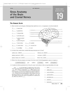
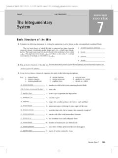
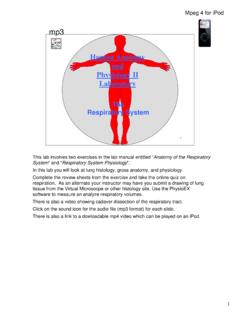
6 Vestibule o 25. serous lining of the abdominal cavity wall y. villi j 26. principal site for the synthesis of vitamin K by microorganisms a z. visceral peritoneum 27. region containing two sphincters through which feces are expelled g 28. bone-supported anterosuperior boundary of the oral cavity 296 Review Sheet 38. ighapmLre38pg295_300 5/12/04 3:25 PM Page 297 impos03 302:bjighapmL:ighapmLrevshts:layouts: 7. Correctly identify all organs depicted in the diagram below. Oral cavity proper Parotid gland and duct Vestibule Pharynx Sublingual gland and ducts Submandibular gland and duct Esophagus Gallbladder Cardiac region of the stomach Liver Pyloric portion of the stomach Hepatic duct Cystic duct Common bile duct Splenic flexure Duodenum (left colic flexure).
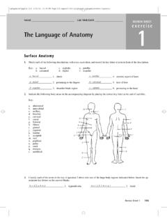
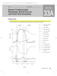
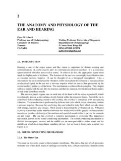
7 Pancreatic duct Pancreas with duct Hepatic flexure (right colic flexure) Transverse colon Jejunum Descending colon Ascending colon Sigmoid colon Ileum Rectum Ileocecal junction Cecum Anal sphincters (Anal canal). Appendix Anus Review Sheet 38 297. ighapmLre38pg295_300 5/12/04 3:25 PM Page 298 impos03 302:bjighapmL:ighapmLrevshts:layouts: 8. You have studied the histological structure of a number of organs in this laboratory. Three of these are diagrammed below. Identify and correctly label each. gastric pit simple columnar epithelium lamina propria villi villi gastric gland intestinal gland Peyer's duodenal gland patches (a) stomach (b) ileum (small intestine) (c) duodenum (small intestine). Accessory Digestive Organs 9.
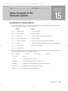
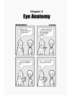
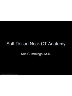
8 Correctly label all structures provided with leader lines in the diagram of a molar below. (Note: Some of the terms in the key for item 10 may be helpful in this task.). Enamel Dentin Crown Pulp cavity Gingiva Neck a Peridontal ligament f Bone Root e Cementum d Root canal b Blood c vessels and nerves in pulp 298 Review Sheet 38. ighapmLre38pg295_300 5/12/04 3:25 PM Page 299 impos03 302:bjighapmL:ighapmLrevshts:layouts: 10. Use the key to identify each tooth area described below. Key: a. anatomical crown c 1. visible portion of the tooth in situ b. cementum b 2. material covering the tooth root c. clinical crown e 3. hardest substance in the body d. dentin h 4. attaches the tooth to bone and surrounding alveolar structures e.
9 Enamel j 5. portion of the tooth embedded in bone f. gingiva d 6. forms the major portion of tooth structure; similar to bone g. odontoblast g 7. produces the dentin h. periodontal ligament i 8. site of blood vessels, nerves, and lymphatics i. pulp a 9. entire portion of the tooth covered with enamel j. root 11. In the human, the number of deciduous teeth is 20 ; the number of permanent teeth is 32 . 2, 1, 2, 3. 12. The dental formula for permanent teeth is 2 32. 2, 1, 2, 3. Explain what this means. There are 2 incisors, 1 canine, 2 premolars, and 3 molars in each jaw (upper and lower) from the median line posteriorly. 2, 1, 0, 2 2I, 1C, 2M. 2 or 2 (no premolars). What is the dental formula for the deciduous teeth?
10 2, 1, 0, 2 2I, 1C, 2M. 13. What teeth are the wisdom teeth ? The number 3 (most posterior) molars. 14. Various types of glands form a part of the alimentary tube wall or duct their secretions into it. Match the glands listed in col- umn B with the function/locations described in column A. Column A Column B. a 1. produce(s) mucus; found in the submucosa of the small intestine a. duodenal glands f 2. produce(s) a product containing amylase that begins starch b. gastric glands breakdown in the mouth e c. intestinal crypts 3. produce(s) a whole spectrum of enzymes and an alkaline fluid that is secreted into the duodenum d. liver d 4. produce(s) bile that it secretes into the duodenum via the bile duct e.