Transcription of Anatomy of the Heart - apchute.com
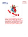
1 NAME _____LAB TIME/DATE _____REVIEW SHEET exercise30 Anatomy of the HeartReview Sheet 30251 Gross Anatomy of the Human anterior view of the Heart is shown here. Match each structure listed on the left with the correct key letter:1. right atrium2. right ventricle3. left atrium4. left ventricle5. superior vena cava6. inferior vena cava7. ascending aorta8. aortic arch9. brachiocephalic artery10. left common carotid subclavian artery12. pulmonary trunk13. right pulmonary artery14. left pulmonary artery15.
2 Ligamentum arteriosum20. left coronary artery16. right pulmonary veins21. circumflex artery17. left pulmonary veins22. anterior interventricular artery18. right coronary artery23. apex of heart19. anterior cardiac vein24. great cardiac veinNAME _____LAB TIME/DATE _____abcdefghijklmnoprstuwvxqgjrubkdnalm ecpofqhitswxvighapmLre30pg251_256 5/12/04 2:47 PM Page 251 impos03 302:bjighapmL:ighapmLrevshts: is the function of the fluid that fills the pericardial sac? the terms in the key to the descriptions provided location of the Heart in the thorax2.
3 Superior Heart chambers3. inferior Heart chambers4. visceral pericardium5. anterooms of the heart6. equals cardiac muscle7. provide nutrient blood to the Heart muscle8. lining of the Heart chambers9. actual pumps of the heart10. drains blood into the right is the function of the valves found in the Heart ? the Heart function with leaky valves? (Think! Can a water pump function with leaky valves?) is the role of the chordae tendineae? :angina pectoris pericarditis 252 Review Sheet 30 reduce friction during Heart enforce a one-way flow of blood through the sThey anchor the AV valve flaps during ventricular systole, thus preventing backflow ofblood into the pain that occurs when the myocardium has insufficient of the 5/12/04 2:47 PM Page 252 impos03 302:bjighapmL:ighapmLrevshts:layouts:Pul monary, Systemic, and Cardiac schematic of a so-called general circulation is shown below.
4 What part of the circulation is missing from this diagram? Add to the diagram as best you can to make it depict a complete systemic/pulmonary circulation and reidentify general cir-culation as the correct clearly between the roles of the pulmonary and systemic circulations. the following scheme of circulation of a red blood cell in the human body:Right atrium through the tricuspid valve to the through the valve to the pulmonary trunk to the to the capillarybeds of the lungs to the to the of the Heart throughthe valve to the through the valve to the to the systemic arteries to the of the tissues to the systemic veins to the ,, andentering the right atrium of the the mitral valve does not close properly, which circulation is affected?
5 Might a thrombus (blood clot) in the anterior descending branch of the left coronary artery cause sudden death? CapillariesArteriesVeinsHeartGeneral circulationReview Sheet 30253 Pulmonary circulation is not distinct from systemic pulmonary circuit provides for gasexchange only; the systemic circuit provides the functional supply of the body ventriclepulmonarysemilunarright and left pulmonary arteriespulmonary veinsleft atriummitral/bicuspidleft ventricleaortic semilunaraortacapillary bedsinferior vena cavasuperior vena cavaSystemicThis artery supplies blood to the interventricular septum and the anterior walls of both ventricles.
6 Ventricular damage, particularly tothe left ventricle, is very arteriesSystemicSystemicSystemic circulationPulmonary capillariesSystemiccapillariesPulmonary veinsighapmLre30pg251_256 5/12/04 2:47 PM Page 253 impos03 302:bjighapmL:ighapmLrevshts:layouts:Mic roscopic Anatomy of Cardiac would you distinguish the structure of cardiac muscle from the structure of skeletal muscle? the following terms to the photoof cardiac muscle at the of cardiac muscle role does the unique structure of cardiac muscle play in its function?
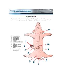
7 (Note: Before attempting a response, describe the unique Anatomy .) Dissection of the Sheep the sheep Heart dissection, you were asked initially to identify the right and left ventricles without cutting into theheart. During this procedure, what differences did you observe between the two chambers?254 Review Sheet 30 Both tissue types are stri-ated; thus, this is not a distinguishing feature. Skeletal muscle cells are long cylindrical cells with many nuclei per cell. Cardiac cellshave one (or two) centrally located nuclei per cell; their branched ends fit together at tight junctions called intercalated discs, whichare not seen in skeletal muscle cells form a functional syncytium by virtue of their intercalated discs.
8 This structural feature plusthe special arrangement of cardiac muscle in the Heart allows the pumping action of the Heart to be carefully coordinated for left ventricle was firmer, thicker, and less compressible; the right ventricle felt flabby. acbdighapmLre30pg251_256 5/12/04 2:47 PM Page 254 impos03 302:bjighapmL:ighapmLrevshts:layouts:Kno wing that structure and function are related, how would you say this structural difference reflects the relative functions of these two Heart chambers? valves prevent backflow into the ; AV valves prevent backflow into the.
9 Using your own observations, explain how the operation of the semilunar valves differs from that of the AV valves. clearly between the location and appearance of pectinate muscle and trabeculae carneae. and contrast the structure of the right and left atrioventricular valves. remnants of fetal structures are observable in the Heart the ligamentum arteriosum and the fossa ovalis. What werethey called in the fetal Heart , where was each located, and what common purpose did they serve as functioning fetal struc-tures?
10 Review Sheet 30255 The left ventricle pumps blood through the high-resistance systemic circulation; therefore, it has to bestronger than the right ventricle, which pumps blood through the short low-resistance pulmonary the ventricle was compressed (as in systole), the AV valve flaps moved superiorly into theclosed position. When water was poured (as when blood backflows) into the semilunar valves, the cusps filled and closed the comblike muscle ridges in the atria. Trabeculae carneae pitted, ridged muscle bundles in the ventricular have thin flaps secured to papillary mus-cles by chordae tendinea.