Transcription of Anterior Segment Surgery and Complications - Mosby
1 10. Anterior Segment Surgery and Complications CATARACT EXTRACTION AND INTRAOCULAR LENS IMPLANTATION. Complications PENETRATING KERATOPLASTY. Complications Correction of Astigmatism in a Corneal Graft LAMELLAR KERATOPLASTY. SUPERFICIAL KERATECTOMY. EXCIMER LASER PHOTOTHERAPEUTIC KERATECTOMY. CONJUNCTIVAL FLAP. LIMBAL STEM CELL TRANSPLANTATION. PTERYGIUM EXCISION AND CONJUNCTIVAL AUTOGRAFT. CONJUNCTIVAL AND CORNEAL TUMOR EXCISION. CORNEAL PERFORATION Surgery . PERMANENT KERATOPROSTHESIS. REFRACTIVE Surgery . Radial Keratotomy Excimer Laser Photorefractive Keratectomy Laser In Situ Keratomileusis CONCLUSION. Anterior Segment Surgery ranges from routine cataract extraction and lens implantation, one of the most common surgical operations in the United States, to rarely performed Surgery such as permanent keratoprosthesis. It also encompasses Surgery first performed centuries ago, such as rudimentary pterygium excision, to the latest in keratorefractive Surgery .
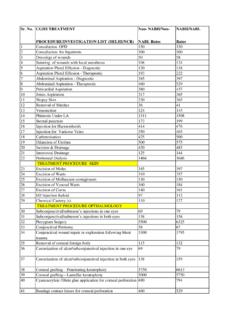
2 CATARACT EXTRACTION AND INTRAOCULAR LENS IMPLANTATION. The many reasons for the development of cataracts are discussed in detail in Chapter 8. Most cataracts are acquired, but they can also be congenital. This section focuses primarily on the treatment of acquired cataracts in adults. Cataracts in adults are generally age related, but some lens opacities may result from other causes such as trauma, inflammation, systemic illness such as diabetes, or medications such as corticosteroids. Cataracts generally advance slowly over years but can advance rapidly over months, or even faster in some patients. The primary indication for cataract extraction is diminished vision caused by the cataract, significantly affecting the patient's lifestyle. The exact point at which this hardship occurs depends on the patient. Certain patients require little visual function and may delay cataract Surgery for years or indefinitely. Other patients with high visual needs seek cataract Surgery with much smaller degrees of visual loss.
3 Rarely, a cataract requires removal because it causes inflammation (phacotoxic uveitis) or elevated intraocular pressure (phacolytic glaucoma). Equally as rare, a cataract may expand to a size large enough to block the outflow of aqueous and cause the intraocular pressure to rise dramatically (phacomorphic glaucoma), also requiring surgical removal. When cataract Surgery is performed, preoperative medications are used for antibiotic prophylaxis and pupillary dilatation. Cycloplegic and mydriatic agents are used in combination to achieve maximal dilation. Nonsteroidal antiinflammatory drugs (NSAIDs) can be used to maintain pupil dilatation during Surgery by blocking prostaglandin- induced miosis resulting from iris manipulation. Anesthesia may include a peribulbar or retrobulbar block and occasionally a facial block. Newer techniques allow topical and intraocular anesthesia to be used in some patients. Such techniques are especially useful in patients using systemic anticoagulants and those with significant systemic disease.
4 Just before Surgery , an antiseptic preparation with povidone-iodine is applied to the eyelids and periocular skin as well as to the conjunctival fornices. The operative eye is then draped in a sterile fashion, and an eyelid speculum is inserted. The most common method to remove a cataract is with an extracapsular technique. This operation may be performed through large or small incisions. The site of the incision is near the limbus, usually superiorly or temporally, but it can be at any location. The basic steps are to make an incision in the eye, open the capsular bag, remove the cataractous lens, and place an intraocular lens implant in the capsular bag. If required, the incision is then sutured closed. The planned extracapsular cataract extraction technique using a superior approach will be described (Fig. 10-1. and Box 10-1). It may involve placement of a bridle suture ( , 4-0 silk) around the superior rectus muscle to assist with exposure of the surgical wound.
5 A superior limbal conjunctival peritomy is performed for approximately 5 clock hours. Hemostasis is achieved with light cautery. A partial-thickness scleral groove may be created just posterior to the surgical limbus. A small scleral tunnel can be made into peripheral clear cornea. The Anterior chamber is entered through the scleral tunnel with a small blade. Viscoelastic is then injected into the Anterior chamber to maintain its depth. A cystotome ( , a bent 25-gauge needle) is placed in the eye and used to create an Anterior capsulotomy. The can-opener technique involves using the cystotome to make multiple small punctures in the Anterior capsule, near the edge of the pupil, in a circular area measuring approximately 5 to 6 mm in diameter. These punctures must be connected so the central circle of Anterior capsule is completely free and can be removed with forceps. The lens nucleus may then be rocked gently with the cystotome to break the nucleus-cortex connections.
6 The cystotome is removed, and corneoscleral scissors or a blade is used to open the wound for approximately 4 to 5 clock hours. Once opened, gentle external pressure is placed a few millimeters behind the limbus inferiorly and superiorly, and the nucleus is slowly expressed from the eye. Once the nucleus is removed, the Anterior chamber is reformed with balanced salt solution. The wound is partially closed with sutures ( , 10-0 nylon). The residual cortex is removed with irrigation and aspiration. This procedure may be performed using manual or automated techniques. Care must be taken to avoid breaking the posterior capsule, which often results in vitreous prolapse and necessitates Anterior vitrectomy. After the cortical material is removed, the capsular bag is filled with viscoelastic, and a posterior chamber lens implant is inserted into the eye and either placed or dialed into the bag. The diameter of the lens optic is generally 6 to 7 mm.
7 The wound is closed with additional sutures as needed, and the viscoelastic is removed with irrigation/aspiration. A miotic may be instilled into the Anterior chamber. The bridle suture is removed, and the conjunctiva is brought down to cover the wound. Subconjunctival or topical antibiotics and corticosteroids are typically used at the conclusion of Surgery (Fig. 10-2). An alternative technique to remove the lens nucleus is phacoemulsification (Fig. 10-3). In this method an ultrasonic probe is used to break up the lens nucleus into small fragments and to remove each piece individually. Because the entire nucleus does not have to exit through the capsular opening in one piece, the opening can be small. A. tear capsulotomy technique (capsulorhexis) was developed to create a continuous circular opening without breaks; it results in a lower tendency to tear posteriorly than the can-opener technique. In the tear capsulotomy method, a break is created in the Anterior capsule with a cystotome, and then the edge of the break is grasped and slowly moved in a circular fashion, connecting at the initial break and creating a 4- to 6-mm diameter opening.
8 The nucleus can then be separated from its epinucleus (hydrodelineation), and the cortex can be separated from the capsule (hydrodissection) by injecting saline through a blunt cannula tip into various portions of the cataractous lens. Many effective methods to remove the lens nucleus have been developed, including bowling, divide-and- conquer, chopping, stop-chop, and supracapsular techniques. Whichever technique is used, the phacoemulsification procedure enables the surgeon to remove the cataract through a small ( to 3-mm) incision. Before the advent of foldable intraocular lenses, this small incision had to be enlarged to allow the lens implant to fit into the eye. Currently, small incision intraocular lenses can be inserted through incisions as small as 2 to 3 mm. The optics of such lenses typically enlarge to 5 to 6 mm in diameter. An incision this small is often self-sealing, averting the need for sutures. Such small incisions have allowed surgeons to move the location of the incision to various clock hours around the eye and also into clear cornea.
9 Many surgeons find that a temporal clear cornea approach works best. With a clear cornea technique, topical anesthetic drops alone can provide sufficient anesthesia for Surgery in selected patients. One percent nonpreserved lidocaine ( ml) can be used intracamerally to supplement topical anesthesia (Box 10-2). Another method to remove cataracts is the intracapsular technique. This operation, popular in the past, is performed infrequently today. It is, however, extremely useful in certain conditions, namely those in which the cataractous lens is subluxed because of broken zonular attachments. In this technique, large (approximately 200 ). conjunctival peritomies and limbal incisions are created, typically in the superior quadrant. An enzyme ( , alpha- chymotrypsin) is instilled into the Anterior chamber to dissolve the zonular adhesions. A suture is placed in the superior lip of the cornea and it is elevated. The superior iris is retracted away from the lens.
10 A cryotherapy probe is placed on the lens, which is frozen and gently extracted from the eye. Ideally, the hyaloid face remains intact and no vitreous prolapses into the Anterior chamber. After the wound is partially closed and viscoelastic is placed in the Anterior chamber, an open-loop Anterior chamber intraocular lens implant can be placed in the angle. A peripheral iridectomy is performed, and the wound is sutured closed. The lack of adequate capsular support does not completely prohibit the insertion of a lens implant in the posterior chamber. A posterior chamber lens can be sutured to the iris or the sclera to secure fixation. A nonabsorbable suture, such as 10-0 prolene, must be used. Additionally, complete Anterior vitrectomy, with special attention to removing all the vitreous in the areas of the lens haptics, is required. One advantage of fixation to the iris is no externalized sutures;. one disadvantage is significant iris-lens touch with potential increased inflammation.