Transcription of Anterior Stabilization of the Shoulder: Latarjet …
1 Department of rehabilitation Services Physical Therapy Anterior Stabilization of the shoulder : Latarjet Protocol shoulder instability may be caused from congenital deformity, recurrent overuse activity, and/or traumatic dislocation. Surgical Stabilization of the glenohumeral joint is indicated after conservative treatment fails and recurrent instability/subluxation continues. A number of different surgical procedures may be indicated in this situation, often divided into soft tissue or bony procedures. shoulder Instability Soft Tissue: Surgical reconstruction targeting the glenohumeral joint s soft tissues for shoulder instability, typically involves labral repairs, the most common being the Bankart repair. A Bankart lesion typically occurs from an Anterior -inferior dislocation of the humerus, tearing the labrum from it s attachment to the glenoid, thereby detaching the inferior gleno-humeral ligament (IGHL).
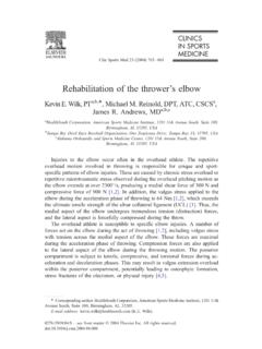
2 Surgical management of this revolves around labral repair to reattach the IGHL under appropriate tension. This may be accomplished either arthroscopically or through an open Most traumatic glenohumeral dislocations may not only cause a Bankart lesion, but may create impression fractures in the postero-superior humeral head termed Hill-Sachs An adverse effect from this procedure includes suturing the capsule too tightly, causing a shortening of the capsule, and thus decreasing the external rotation allowed at the glenohumeral joint. Other complications are extremely rare, but may include axillary nerve damage, subscapularis rupture (seen only in open repairs performed with subscapularis detachment and repair), and recurrent instability. If there is bony deficiency in the glenoid, which represents 20% or more of the antero-inferior glenoid, it is biomechanically impossible to restore the same stability and is therefore more prone to recurrent instability and failure.
3 shoulder Instability Bony Deficiency: In cases where significant bony deficiency is present (where greater than 20% of the glenoid s surface area is missing) addressing only the soft tissue issues during the surgical procedure may lead to eventual recurrence of instability. Bony deficiency can result from congenital deformity, trauma, or recurrent dislocation. These lesions are not well visualized on plain films and are best seen on 3-dimensional CT scan (see Fig. 1). Anterior Stabilization of the shoulder : Latarjet Protocol Copyright 2009 The Brigham and Women's Hospital, Inc. Department of rehabilitation Services. All rights reserved. 1 Figure 1: A 3D CT reconstruction of the scapula. The blue segment illustrates the bony deficit of the glenoid. When bony lesions reach critical dimensions, reconstruction of this deficit using autograft bone yields the best surgical results.
4 The Latarjet procedure is the most popular and highly effective, transferring the distal coracoid into the bony Surgical Technique A deltopectoral approach is used to expose the coracoid process. The corcoacromial ligament and the pectoralis minor attachment are divided, where as coracobrachialis and the short head of the biceps origins remain intact. The coracoid is osteotomized at its knee yielding bony graft approximately cm in length. Great care is taken to avoid damage to the soft tissues and musculotaneous nerve in the surrounding area. With the arm in external rotation, the subscapularis muscle is either split along its length or detached from the lesser tuberosity and the joint is exposed. The graft is shaped and contoured to fill the defect and is secured with screw fixation placed at the antero-inferior glenoid.
5 With the corachobracialis and biceps still attached to the coracoid, they now serve as a dynamic sling further stabilizing the glenohumeral The subscapularis split is then repaired. The final bony reconstruction is illustrated in Figure 2. Anterior Stabilization of the shoulder : Latarjet Protocol Copyright 2009 The Brigham and Women's Hospital, Inc. Department of rehabilitation Services. All rights reserved. 2 Figure 2: Illustration of the coracoid transfer to correct the inferior glenoid bony deficiency. Potential Complications There are several possible complications that could occur after a Latarjet procedure. Considering the coracoid osteotomy, there is a risk for non-union of the transferred coracoid process, which occurs typically in 3% of In a long-term follow-up by Banas et al, 82% had bony union and 14% had fibrous union of the coracoid and glenoid.
6 4 Despite the bony union, many patients continued to experience discomfort years post-operatively and underwent another procedure to extract the screws. Screw loosening and screw breakage are other possible reasons a patient may undergo a screw removal procedure. A follow-up performed by Banas et al, found 14% of shoulders required secondary operations, 4% for Stabilization , and 10% for screw removal secondary to discomfort. Current research is evaluating the optimal screw placement during the procedure to reduce loosening, breakage, and Other complications, including musculocutaneous nerve palsy and subscapularis dysfunction, are reported but rare events. Following a Latarjet procedure, the most functional limitation reported is a decrease in external rotation range of motion. Although some patients may return to overhead throwing sports, most do not regain full external According to Hovelius and colleagues, the mean loss of external rotation was degrees in adduction and 8 degrees in abduction.
7 The complications of rotator cuff tendonitis and limitation in external rotation can be reduced with proper progression in Anterior Stabilization of the shoulder : Latarjet Protocol Copyright 2009 The Brigham and Women's Hospital, Inc. Department of rehabilitation Services. All rights reserved. 3 Anterior Stabilization of the shoulder : Latarjet Protocol Copyright 2009 The Brigham and Women's Hospital, Inc. Department of rehabilitation Services. All rights reserved. 4 rehabilitation Considerations One must recall that the purpose of the Latarjet procedure is to reinstate Anterior stability to the glenohumeral joint. While this is primarily a bony procedure, specific attention must be directed towards the soft tissues which play a critical role in maintaining stability. Early post-operative therapy must protect the repair of the subscapularis as well as the developing bony union of the coracoid process.
8 Since it will take approximately 6-8 weeks to form an osseous union of the newly reconstructed glenoid, the biceps and coracobrachialis attachment to the coracoid needs to be protected during the initial postoperative period. Aggressive shoulder extension and combined extension and external rotation stretching is not indicated. Once strengthening commences, a gradual progressed program of biceps and coracobrachialis strengthening needs to be followed to minimize undue stress and tension on their muscular origins. In addition, isolated external rotation range of motion needs to be gradually regained after surgery to allow the Anterior capsule and subscapularis to heal For that reason, external rotation range of motion is advanced in a protected fashion, with early emphasis on external rotation work being done in an open packed position ( scapular plane at about 45 degrees of abduction) and then progressed to positions that gradually tension the subscapularis ( full adduction and then at 90 degrees of abduction and above).
9 Please refer to protocol below for more detail. (In the case of a subscapularis take down and repair, external rotation gains need to be progressed slower and one should avoid aggressive external rotation stretching and internal rotation strengthening until the subscapularis is well healed. In these cases it is helpful to get a safe zone of initial external rotation range of motion from the referring surgeon, as determined from intraoperative inspection from either the operative note or discussion with surgeon.) Due to the surgical technique and early immobilization required to promote healing, the subscapularis may not only be impacted in terms of length, but in terms of force production and proprioception. Hence, specific subscapularis proprioception and strengthening needs to be incorporated to enhance subscapularis function postoperatively.
10 The clinician needs to tailor the rehabilitation program to address the unique structure of the subscapularis to enhance both the upper and lower subscapularis fibers. This is warranted due to the fact that the subscapularis is innervated by both the upper and lower subscapular nerves, along with the presence of two different muscular fiber alignments; hence, its action has been described as being like that of two different muscles depending upon the functional The upper fibers are primarily aligned in a horizontal fashion and the lower fibers are arranged in more of an oblique alignment. One must therefore be selective in the rehabilitation protocol to maximally stimulate the appropriate portion of the subscapularis with the correct exercise. Anterior Stabilization of the shoulder : Latarjet Protocol Copyright 2009 The Brigham and Women's Hospital, Inc.