Transcription of APPLICATIONS PRINCIPLES AND CLINICAL MONITORING ...
1 INVASIVE HEMODYNAMICMONITORING: PHYSIOLOGICALPRINCIPLES AND CLINICALAPPLICATIONSE dwards Lifesciences, Edwards, the stylized E logo, CCOmbo V,STAT and VIP are trademarks of Edwards LifesciencesCorporation. CCOmbo, Swan-Ganz and Vigilance are trade-marks of Edwards Lifesciences Corporation and are registeredin the Patent and Trademark :Please refer to current product pakage inserts forindication, contraindictions, warning, precautions, and instruc-tions for use. Copyright 2002 Edwards Lifesciences LLCAll rights reserved. 1147-7/00-CCDepartment of Professional EducationProviding 25 years of excellence in critical care education Consultation Educational Programs and Materials 24-Hour CLINICAL and Technical Ext. 2677 Technical Assistance HotlineOur 24-Hour Technical Assistance Hotline isdedicated to quick problem ServiceOur customer service department is availablefrom 6:00 to 4:30 ( ) for orderentry or product information.
2 We are alsoproud to offer 24-Hour service for emergencysituations during our Lifesciences LLC One Edwards Way Irvine, CA 92614 Phone: Lifesciences (Canada) Inc. 4 Robert Speck Parkway, Suite 900 Mississauga, Ontario Canada L4Z 1S1 Phone: Lifesciences Au Glapin 1162 Saint-Prex Switzerland Phone: Lifesciences Japan 2-8 Rokubancho Chiyoda-ku Tokyo 102-0085 Japan Phone: 81-3-5213-5700 INVASIVEHEMODYNAMICMONITORING:PHYSIOLOGI CALPRINCIPLES ANDCLINICAL APPLICATIONSA uthor: Jan M. Headley, RN, BS, CCRNI rvine, CaliforniaTABLE OF CONTENTSI ntroduction ..1 functional anatomy ..2 Right vs. Left Heart ..2 Cardiac Cycle ..3 Systole ..3 Diastole ..4 Applicable Cardiac Physiology ..5 Cardiac Output ..5 Determinants of Cardiac Output ..5 Heart Rate ..5 Stroke Volume.
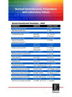
3 5 Frank-Starling Law ..6 Preload ..6 Afterload ..7 Contractility ..8 Summary ..8 Myocardial Oxygen Consumption ..9 Physiological Basis of Pulmonary ArteryPressure MONITORING ..10 Insertion Techniques for the Swan-Ganz Catheter ..12 Continuous Pressure MONITORING ..14 Summary Guidelines for Safe Use of Balloon-Tipped Pulmonary Artery Catheters ..16 Cardiac Output Determinations ..18 The Fick Method ..18 Dye Indicator Dilution Method ..18 Thermodilution Method ..19 CLINICAL APPLICATIONS ..21 Evaluation of Cardiac Performance ..21 Significance of Hemodynamic Measurements ..21 Direct Measurements ..21 Derived Parameters ..22 Utilizing the Swan-Ganz Catheter for Differential Diagnosis ..24 Low Output States ..24 Limitations of Hemodynamic MONITORING ..26 Left Ventricular End Diastolic Volumes vs.
4 End Diastolic Pressure ..26 Pulmonary Artery Wedge vs. Left Ventricular End Diastolic Pressures ..27 Conditions in Which PAW is Greater Than LVEDP ..27 Conditions in Which PAD is Less Than LVEDP ..27 Conditions in Which PAD Does Not Equal PAW (1 - 4 mm Hg) ..27 Complications ..28 Intra-Arterial MONITORING ..29 Components of the Arterial Pulse ..29 Mean Arterial Pressure ..30 Arterial Waveforms for Differential Diagnosis ..31 Summary ..33 Bibliography ..34 Appendix I ..36 INTRODUCTIOND uring the last 25 years, the art of critical care medicine hasgreatly changed. This process has been due in part to theformation of specialized units for patient care, advances intechnology, and a better understanding of physiology byhealth care of the earlier advances in technology that helped todrive this progress was the development in the 1960 s of theEdwards Swan-Ganz catheter.
5 In the early 1970 s, theaddition of a thermistor to the catheter allowed for rapidassessment of cardiac output. At the same time, moresophisticated MONITORING systems were also being a result, more complete hemodynamic assessment couldbe carried out with relative ease at the patient s advanced technology comes the requirement ofadvanced clinicians. The critical care practitioner must bebetter educated in order to practice effectively. The goal ofthis booklet is to provide the practitioner with an expandedknowledge of basic hemodynamic book is divided into sections that discuss the variouscomponents of hemodynamic MONITORING , including: functional anatomy and applicable cardiac physiology,physiological bases of hemodynamic MONITORING , cardiacoutput determinations, and CLINICAL APPLICATIONS . The bookmay be read as a whole or each section referred to as a single ANATOMYR ight vs.
6 Left HeartIn this section, a review of functional cardiac anatomy andapplicable physiology is presented. These concepts providethe foundations of hemodynamic discussing the functional anatomy of the heart inregards to hemodynamic MONITORING , the heart is describedas two separate pumps. Each side, or pump, has its ownfunction and pressure generation. For this reason, the termsright and left heart are as a single unit, the right heart consists of the rightatrium and right ventricle. The right heart s main function isto receive deoxygenated venous blood into the right there, the right ventricle needs to generate only aminimal amount of pressure to pump the blood through thepulmonic valve into the pulmonary circulation. It is becauseof this that the right heart is considered a low left heart is a similar unit which receives oxygenatedblood from the pulmonary system.
7 The left heart isconsidered a high pressure system since the left ventricleneeds to generate a greater amount of pressure to pumpblood through the aortic valve, into the aorta, and thenthrough the systemic pulmonary capillary bed lies between the right and leftheart. The capillary bed is a very compliant system with ahigh capacity to hold blood. Changes in this capacity can bedetected through pressure changes seen for both sides of circulatory system therefore consists of two circuits in aseries: pulmonic circulation, which is a low-pressure systemwith low resistance to blood flow; and the systemiccirculation, which is a high-pressure system with highresistance to blood events are classified as either systolic ordiastolic. By convention, the terms usually depict ventricularactivity. The atria have phases of systole and diastole as dothe ventricles.
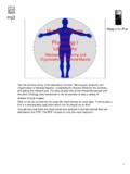
8 Technically speaking, the left side of the heartis the first to begin and complete the systolic and diastolicphases. For ease of discussion and since the time differenceis minimal, we will consider the right and left sides tofunction at the same time. Figure 1 Anatomical Structure Location and AbbreviationsRight HeartLeft HeartSVC - Superior Vena CavaLA - Left AtriumIVC - Inferior Vena CavaLV - Left VentricleRA - Right AtriumAo - AortaRV - Right VentricleMV - Mitral ValveTV - Tricuspid ValveAoV - Aortic ValvePV - Pulmonic ValvePA - Pulmonary Artery2 CARDIAC CYCLEThe cardiac cycle consists of nearly synchronized activity ofthe atria and ventricles. The sequence is essentially the samefor the right and left sides of the heart. For generaldiscussion, systole and diastole are the two basic , when examining the cycles closer there are manydifferent sub-phases for both.
9 The purpose of this section isto discuss the important phases of the cardiac , the ECG has been the basis for noting systoleand diastole. For more precise identification of intracardiacwaveforms, the delineation of electrical versus mechanicalcardiac cycle is addressed first type of cardiac cycle that must occur is theelectrical cardiac cycle. The initial phase is depolarization,which begins from the sinus node and spreads a wave ofelectrical current throughout the atria. This current is thentransmitted throughout the ventricles. Following the wave ofdepolarization, muscle fibers contract, which next electrical activity is repolarization which results inthe relaxation of the muscle fibers and produces diastole. Inthe normal heart, initial electrical activity produces themechanical activity of systole and diastole.
10 There is a timedifference between the two called electro-mechanicalcoupling, or the excitation-contraction phase. When lookingat a simultaneous recording of the electrocardiogram andpressure tracing, the ECG will show the appropriate wavebefore the mechanical tracings previously mentioned, systole and diastole are generallyused in relation to ventricular activity, since it is theventricles that are responsible for performing the pumpingaction. It is important to remember that while the ventriclesare in systole, the atria are in diastole, and that while theventricles are in diastole, the atria are in systole. This willbecome more apparent as we go through the various phasesof the cardiac cardiac cycle is a continuous cycle of pressure changesand blood flow. It doesn t matter if the discussion beginswith systole or diastole.