Transcription of Applied anatomy of the wrist, thumb and hand
1 Copyright 2013 Elsevier, Ltd. All rights anatomy of the wrist, thumb and hand CHAPTER CONTENTSJ oints, joint capsules and ligaments .. e102 Distal radioulnar joint ..e102 Wrist joint ..e103 Carpometacarpal joints ..e104 Trapezium first metacarpal joint ..e104 Metacarpophalangeal joints ..e105 Interphalangeal joints ..e105 Muscles and tendons .. e105 Extrinsic muscles and tendons ..e105 Intrinsic muscles..e107 Nerve structures .. e109 Median nerve ..e109 Ulnar nerve ..e110 radial nerve ..e111 The anatomy of wrist, thumb and hand is complex because of the presence of many different functional joints: the distal radioulnar joint, the wrist joint (containing the radiocarpal and the intercarpal joints, the carpometacarpal joints, the trapezium first metacarpal joint), the metacarpophalangeal joints and the interphalangeal joints.
2 The contractile structures can be divided into extrinsic and intrinsic muscles. The former the long tendons are quite regularly affected, while the latter are clinically of less , joint capsules and ligamentsDistal radioulnar jointThe joint is a uniaxial pivot joint. The articular surfaces are between the convex head of the ulna and the concave ulnar notch of the radius. A fibrocartilaginous articular disc binds the distal end of the ulnar notch on the radius to the ulnar styloid process. This disc is part of the so-called triangular fibrocarti-lage complex (TFCC) and sits between the ulnar head and the ulnar carpus (lunate and triquetrum).
3 Therefore the distal radioulnar joint can be seen as L-shaped: the short leg is between radius and ulna and the longer leg between the distal ulna and the articular disc (see Putz, Fig. 318). It has a loose capsule, reinforced with some ligaments, that allows rotation movement of the radius about the ulna. The two bones are held together mainly by the triangular fibrocartilage complex (TFCC) and to a lesser degree by the interosseous membrane and the pronator quadratus muscle (Fig. 1).The TFCC contains different parts: (a) the triangular fibro-cartilage, which has a central component the articular disc and two adjoining ligaments, the dorsal and palmar radioulnar ligaments.
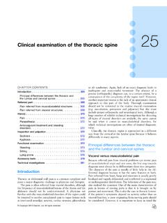
4 These take their origin at the dorsal and volar edges of the sigmoid notch and insert onto the ulnar styloid base (b) a meniscus homologue. This thickened ulnar part of the TFCC, inserts into the triquetral and hamate bones and the base of the fifth metacarpal; (c) the ulnar collateral ligament; (d) the ulnolunate and ulnotriquetral ligaments; and (e) the sheath of the extensor carpi ulnaris tendon, which is strongly attached to the posterolateral aspect of the TFCC. The TFCC stabilizes the radioulnar and ulnocarpal joints, transmits and distributes load from the carpus to the ulna and facilitates complex movements to the wrist (Fig.)
5 2).This distal radioulnar joint allows pronation supination movements of the forearm, inevitably in combination with movement at the upper radioulnar joint, around an axis which runs through the head of the ulna. The distal end of the radius makes a circumferential gliding movement around and in front of the head of the ulna (see online chapter Applied anatomy of the elbow). Pronation has an amplitude of 85 and is stopped by the radius impacting against the ulna and by tension in the dorsal radioulnar ligament and the interosseous mem-brane.
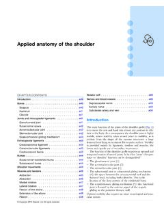
6 Consequently, there is an elastic end-feel. Supination ceases at an angle of 90 when the posterior aspect of the ulnar Applied anatomy of the wrist, thumb and hand e103 Copyright 2013 Elsevier, Ltd. All rights , triquetral and pisiform bones, and the distal row the trapezium, trapezoid, capitate and hamate (Fig. 3, see Stand-ring, Fig. ).The wrist joint is complex: there are two components, proximal and distal (see Putz, Fig. 326). Proximally the distal part of the radius and the articular disc articulate with the proximal row of carpal bones to form a condylar joint (radio-carpal joint) which has its concave surface at the radial side.
7 The joint moves along two axes: anteroposterior for ulnar and radial deviation, and transverse for flexion and extension (Fig. 4).Distally, the intercarpal joint is between the proximal and distal rows of bones (Fig. 5), is an open S-shape and acts as a hinge. It should not be considered as an independent joint because its function is to augment the mobility of the carpal bones and thus allow greater mobility at the are restricted by collateral (Fig. 5), palmar and dorsal ligaments (Fig. 6). radial and ulnar collateral ligamentsThe radial collateral ligament, between the styloid process of the radius and the scaphoid bone, is taut when the hand is brought into ulnar deviation.
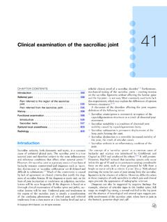
8 The ulnar collateral ligament, between the styloid process of the ulna and the triquetral and pisiform bones, is under tension during radial deviation of the ligamentsThe dorsal wrist ligaments are comparatively thin. They are reinforced by the floor and septa of the fibrous tunnels for the six dorsal compartments (see below) and have a Z-shaped configuration. The fibres of the dorsal radiocarpal ligaments are aligned more or less in the same axis as the forearm, those of Fig 1 Right distal radioulnar joint in supination.
9 T, triquetrum; H, hamate; L, lunate; S, scaphoid; U, ulna; UCL, ulnocapitate ligament. TULSHUCLFig 2 Right distal radioulnar joint in pronation. Ulnolunate ligamentUlnotriquetralligamentArticular discUlnaRadiusLunateTriquetrumMeniscus homologueDorsal radioulnarligamentPalmar radioulnarligamentExtensor carpiulnaris sheathFig 3 Palmar carpus of the left hand. From Standring, Gray s anatomy , 40th edn. Churchill Livingstone/Elsevier, Philadelphia, 2009 with ofhamateCapitatePisiformTriquetrumLunate TrapezoidTrapeziumTubercle of scaphoidMetacarpalsnotch of the radius is brought into contact with the styloid process of the ulna through the extensor carpi ulnaris tendon.
10 Its end-feel is also elastic (see Standring, Fig. ).Wrist jointThe wrist joint consists of two rows of carpal bones. The proximal row contains (from radius to ulna) the scaphoid, The Wrist, thumb and Hande104 Copyright 2013 Elsevier, Ltd. All rights These palmar ligaments are of little clinical importance and are taut during extension of the amplitude of radial deviation is only 15 , whereas ulnar deviation has an amplitude of approximately 45 , the wrist being held in the neutral position between flexion and exten-sion.