Transcription of Approach to Respiratory Distress in Dogs & Cats
1 | November/December 2015 | TODAY S VETERINARY PRACTICEAPPROACH TO Respiratory DISTRESSPeer Reviewed 53 Managing dogs and cats in Respiratory Distress is a multifaceted effort that involves stabilizing patients prior to determining a defi nitive diagnosis. Fortunately, Respiratory Distress no matter what the cause requires somewhat standardized interventions during initial stabilization. INITIAL STABILIZATIONOne of the benefits of initial stabilization is that it provides the practitioner time to consider the appropriate diagnostic and subsequent therapeutic SupplementationInitial stabilization of a patient in Respiratory Distress generally involves provision of oxygen supplementation, with or without patient sedation. The most common type of oxygen supplementation provided is use of an oxygen cage with a high fraction of inspired oxygen (FiO2) (eg, 40% 60%); a face mask or flow-by oxygen from a hose can also be used. In more extreme cases, animals in Respiratory Distress may require emergency intubation, higher FiO2 (eg, 100%), and provision of positive pressure ventilation in order to provide adequate Respiratory stabilization.
2 Particularly in cases of upper airway obstruction, the practitioner may need to ensure a patent airway by intubation or tracheostomy (if oral intubation is not possible).SedationSedation with careful monitoring and, if necessary, intubation and ventilation can be extremely useful in animals that have become anxious due to hypoxemia and/or hypercapnia. In some patients, especially dogs with upper airway obstruction, stabilization may require sedating the animal by administering some form of anesthetic induction agent; then clearing the oral cavity of obstructing material (eg, secretions or foreign material in a choking animal) prior to intubation or tracheostomy. Cooling MeasuresAnimals with upper airway obstruction, such as those with laryngeal paralysis, may become hyperthermic due to the increased work of breathing. Because of the airway obstruction, these animals are unable to effectively pant, resulting in inability to thermoregulate and dissipate heat.
3 As such, cooling hyperthermic patients in Respiratory Distress is an important component of initial stabilization, and can be accomplished by: Administering room temperature IV fluids Covering the patient with wet towels Putting a fan on the patient Applying alcohol to the axilla, inguinal area, and feet. Active cooling should stop once the patient s temperature reaches 103 F to avoid precipitating hypothermia. ThoracocentesisInitial stabilization may also include thoracocentesis, if severe Respiratory Distress is secondary to pleural space disease, such as pneumothorax or pleural DIAGNOSTIC APPROACHD iagnostic Approach to a patient in Respiratory Distress should consider the patient s signalment and history as well as the broad anatomic differential diagnoses of dyspnea (Table 1, page 54). SignalmentClues in the patient s signalment are common. For example: Upper airway obstruction due to brachycephalic airway disease is a common cause of Respiratory Distress in brachycephalic dogs, such as English bulldogs.
4 Cardiogenic pulmonary edema is a common cause of Respiratory Distress in small breed dogs with chronic valvular disease (eg, mitral endocardiosis), such as Cavalier King Charles spaniels. Lower airway obstruction associated with asthma is a common cause of Respiratory Distress in cats, with certain breeds, such as the Siamese, overrepresented. Approach to Respiratory Distress in Dogs & CatsClaire R. Sharp, BSc, BVMS (Hons), MS, CMAVA, Diplomate ACVECC Tufts UniversityMinimizing Stress dogs and cats with Respiratory Distress are often fragile and can decompensate rapidly. Initial evaluation should be performed rapidly, with minimal stress to the patient. Often, one of the best fi rst steps is to place the animal in an oxygen cage and allow it to relax, considering it has usually been through a stressful car ride and changed environments (home to car to clinic) that can exacerbate Distress . Peer Reviewed 53 Today s VeTerinary PracTice | november/december 2015 | Approach To Respiratory disTressPeer reviewed 54 HistoryHistory can also be extremely useful; for example: History of blunt trauma (eg, hit by a car) should prompt concern for pulmonary contusions, pneumothorax, diaphragmatic hernia, or flail chest.
5 In cats, a history of cough is consistent with asthma, while in dogs, a cough might suggest tracheobronchial disease, interstitial lung disease, or pulmonary edema. physical ExaminationExamining a patient with Respiratory Distress should involve: 1. Initial observation: Consider breathing pattern, presence of externally audible noise with breathing, any signs of trauma, or abdominal distension2. Lung auscultation: Increased adventitial lung sounds (eg, crackles, wheezes, harsh lung sounds) are associated with lower airway and pulmonary parenchymal disease Decreased lung sounds, in an animal with Respiratory Distress , are associated with pleural space disease. 3. Cardiac auscultation: A murmur, gallop, or other arrhythmia may indicate underlying cardiac disease and the potential for cardiogenic pulmonary edema or pleural general, breathing patterns help narrow the list of differential diagnoses (Table 1). For example, upper airway obstruction is associated with inspiratory dyspnea and an externally audible noise.
6 In contrast, lower airway obstruction tends to be associated with expiratory dyspnea and wheeze, with the wheeze generally just audible on thoracic auscultation with a stethoscope, rather than externally TestsExtensive diagnostics should not be performed until the patient has been stabilized as much as possible, a brief physical examination has been performed, and the practitioner has localized the disease to the most likely anatomic location (Table 1). Diagnostic tests may subsequently involve: Blood analysis: Screening blood tests, blood gases Imaging: Thoracic ultrasound, including focused assessment with sonography for trauma, triage, and tracking (tFAST); thoracic radiographs; thoracic computed tomography (CT); or echocardiography Respiratory fluid analysis: Bronchoalveolar lavage, thoracocentesisCategories of Respiratory DiseaseDogs and cats with Respiratory Distress can be classified into 8 disease categories, some of which are associated with distinct breathing patterns observed during physical ,2 These categories include both primary Respiratory diseases and secondary causes of Respiratory difficulty.
7 Diagnostic Approach is determined by the category of disease causing Respiratory 1. Anatomic Classification: Causes of Respiratory DistressDISEASE CATEGORYEXAMPLESBREATHING PATTERN1. Upper Airway Obstruction Brachycephalic airway disease Laryngeal paralysis Inspiratory dyspnea Externally audible noise (eg, stertor, stridor)2. Lower Airway Obstruction Asthma Expiratory dyspnea Wheeze (audible with stethoscope)3. Pulmonary Parenchymal Disease Pneumonia Interstitial lung disease Pulmonary edema Pulmonary contusions Not consistent; may be rapid, shallow, or both, and have both inspiratory and expira-tory components4. Vascular Pulmonary thromboembolism Not specific5. Pleural Space Disease Pneumothorax Pleural effusion Inspiratory dyspnea, rapid shallow breathing, or generalized paradoxical breathing Reduced lung sounds on auscultation6. Flail Chest Focal paradoxical breathing7. Abdominal Distension Ascites Organomegaly Inspiratory dyspnea8.
8 Look-alike Diseases Not | November/December 2015 | ToDay s VeTeriNary PracTice Approach To Respiratory DisTressPeer reviewed 55 Airway examination : Upper airway examination , tracheobronchoscopy Drug trials: Such as bronchodilators, diuretics, and AIRWAY OBSTRUCTIONE tiologyUpper Respiratory tract obstruction involves a mechanical or functional obstruction of the upper (large) airways (ie, the pharynx, larynx, or trachea). Nasal disorders are not considered in this article as the animal should always be able to open its mouth and breathe, preventing the development of dyspnea even if the nasal cavity is obstructed. Specific causes of upper airway obstruction include: Naso-oropharyngeal disorders, including polyps (especially in cats), masses, and foreign bodies Severe head trauma that results in bone fractures (especially nasal, jaw, and palatine fractures) and associated hemorrhage and swelling Laryngeal disorders, including laryngeal paralysis, laryngeal collapse, laryngeal masses (eg, neoplasia, abscesses, granulomas), and laryngeal inflammation Tracheal diseases, including tracheal collapse, tracheal foreign body, tracheal stenosis, stricture, tracheal tear, or tracheal mass (either intra- or extraluminal) Brachycephalic airway disease, which involves a combination of primary and secondary anatomic abnormalities of the upper airways, including stenotic nares, an elongated soft palate, everted laryngeal saccules, laryngeal edema and/or collapse and, in some breeds (eg, English bulldog)
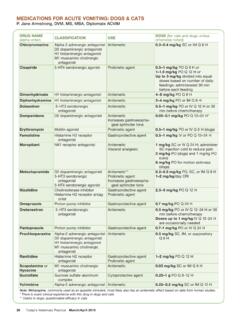
9 , a hypoplastic SignsCharacteristic signs in patients with an upper airway obstruction include inspiratory Distress and an externally audible noise associated with breathing (eg, stertor, stridor). Tracheal disease is usually associated with a StabilizationInitial stabilization and therapy may involve: Oxygen administration/securing an airway: Generally administered by face mask (if tolerated), flow-by oxygen, or oxygen cage, with intubation or tracheostomy performed if needed Sedation: Achieved with anxiolytic drugs, such as acepromazine or dexmedetomidine, or sedative analgesics, such as butorphanol (Table 2) Cooling: Many dogs with upper airway obstructions become hyperthermic due to inability to dissipate heat through their upper airways; the goal is to reduce body temperature to at least 103 F, while avoiding hypothermia Corticosteroids: Breathing against an obstruction can result in marked edema of the upper airway soft tissue; therefore, anti-inflammatory doses of corticosteroids (eg, dexamethasone SP, mg/kg IV single dose or Q 24 H) can be ApproachOnce the patient is stable, diagnostic tests can be pursued.
10 Upper airway examination . examination is performed after preoxygenation under sedation. At its most basic, examination may involve inspection of the oropharynx and larynx with a laryngoscope; in patients with suspected tracheal disease, it Sedation for Patients in Respiratory Distress In dogs, reasonable choices for sedation are butorphanol, acepromazine, or dexmedetomidine, while butorphanol is the drug of choice in cats. Choice of drug(s) used for sedation/anxiolysis should be based on the individual drug s properties, and relative risks versus benefits for the patient. For example: Butorphanol is a very safe and effective drug at recommended doses; however, it is relatively short acting (often only 1 2 hours) and cannot easily be reversed. Acepromazine is also very effective; however, it may be more likely to produce undesirable effects, such as hypotension; has a long duration of action (4 6+ hours); and cannot be reversed. Dexmedetomidine has the desirable quality of being reversible (with atipamezole) and titratable (given a short duration of action); however, it can produce undesirable effects, such as bradycardia and hypotension.