Transcription of ARTICLE IN PRESS - 中山醫學大學附設醫院
1 ReviewUterine sarcomas: A reviewEmanuela D'Angelo, Jaime Prat Department of Pathology, Hospital de la Santa Creu i Sant Pau, Autonomous University of Barcelona, Sant Antoni M. Claret, 167, 08025 Barcelona, Spainabstractarticle infoArticle history:Received 29 June 2009 Available online xxxxKeywords:Uterine sarcomasLeiomyosarcomaEndometrial stromal sarcomaUndifferentiated endometrial sarcomas are rare tumors that account for 3% of uterine cancers. Their histopathologicclassification was revised by the World Health Organization (WHO) in 2003. A new staging system has beenrecently designed by the International Federation of Gynecology and Obstetrics (FIGO). Currently, there is noconsensus on risk factors for adverse outcome. This review summarizes the available clinicopathological dataon uterine sarcomas classified by the WHO diagnostic was searched between 1976 and 2009 for all publications in English where the studiedpopulation included women diagnosed of uterine carcinosarcomas (malignant mixed mesodermal tumors or MMMT) are currently classifiedas metaplastic carcinomas, leiomyosarcomas remain the most common uterine sarcomas.
2 Exclusion ofseveral histologic variants of leiomyoma, as well as smooth muscle tumors of uncertain malignantpotential, frequently misdiagnosed as sarcomas, has made apparent that leiomyosarcomas are associatedwith poor prognosis even when seemingly confined to the uterus. Endometrial stromal sarcomas areindolent tumors associated with long-term survival. Undifferentiated endometrial sarcomas exhibitingnuclear pleomorphism behave more aggressively than tumors showing nuclear uniformity. Adenosarcomashave a favorable prognosis except for tumors showing myometrial invasion or sarcomatous may represent well-differentiated adenosarcomas. The prognosis of carcinosarcomas (whichare considered here in a post-script fashion) is usually worse than that of grade 3 endometrial expression of Ki67, p53, and p16 is significantly higher in leiomyosarcomas andundifferentiated endometrial sarcomas than in endometrial stromal of H&E stained sections has been equivocal in the prediction of behavior ofuterine sarcomas.
3 Immunohistochemical studies of oncoproteins as well as molecular analysis of non-random translocations will undoubtedly lead to an accurate and prognostically relevant classification ofthese rare tumors. 2009 Elsevier Inc. All rights 0 Clinical 0 Pathological 0 Immunohistochemistry and molecular 0 Prognosis and 0 Smooth muscle tumors of uncertain malignant potential (STUMP).. 0 Endometrial stromal 0 Endometrial stromal 0 Low-grade endometrial stromal 0 Immunohistochemistry and molecular 0 Prognosis and 0 Undifferentiated endometrial 0 Clinicopathological 0 Prognosis and 0 Gynecologic Oncology xxx (2009) xxx xxx Corresponding author. Fax: +34 93 291 93 Prat).YGYNO-973334; No. of pages: 9; 4C: 3, 60090-8258/$ see front matter 2009 Elsevier Inc. All rights lists available atScienceDirectGynecologic Oncologyjournal homepage: IN PRESSP lease cite this ARTICLE as: D'Angelo E, Prat J, Uterine sarcomas: A review, Gynecol Oncol (2009), 0 Clinical 0 Pathological 0 Adenosarcoma versus 0 Prognosis and 0 Carcinosarcoma (malignant mixed mullerian tumor).
4 0 Clinical 0 Pathological 0 Prognosis and 0 Conflict of interest 0 IntroductionUterine sarcomas are rare tumors that account for approximately1% of female genital tract malignancies and 3% to 7% of uterine cancers[1]. Although the aggressive behavior of most cases is well recognized,their rarity and histopathological diversity has contributed to thelack of consensus on risk factors for poor outcome and optimal treat-ment[2].Histologically, uterine sarcomas werefirst classified into carcino-sarcomas, accounting for 40% of cases, leiomyosarcomas (40%),endometrial stromal sarcomas (10% to 15%), and undifferentiatedsarcomas (5% to 10%). Recently, carcinosarcoma has been reclassifiedas a dedifferentiated or metaplastic form of endometrial this, and probably because it behaves more aggressively thanthe ordinary endometrial carcinoma, carcinosarcoma is still includedin most retrospective studies of uterine sarcomas, as well as in the2003 World Health Organization (WHO) classification[3].
5 The 1988 International Federation of Gynecology and Obstetrics(FIGO) criteria for endometrial carcinoma have been used until now toassign stages for uterine sarcomas in spite of the different biologicbehavior of both tumor categories. Recently, however, a new FIGO classification and staging system has been specifically designed foruterine sarcomas in an attempt to reflect their different biologicbehavior (Table 1) [4]. Briefly, three new classifications have beendeveloped: (1) staging for leiomyosarcomas and endometrial stromalsarcomas; (2) staging for adenosarcomas; and (3) staging for carcino-sarcomas (MMMT). Whereas in thefirst classification stage I sarcomasare subdivided according to size, subdivision of stage I adenosarcomastakes into account myometrial invasion. On the other hand, carcino-sarcomas will continue to be staged as endometrial featuresAfter excluding carcinosarcoma (MMMT), leiomyosarcoma hasbecome the most common subtype of uterine sarcoma.
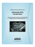
6 However, itaccounts for only 1 2% of uterine malignancies. Most occur in womenover 40 years of age who usually present with abnormal vaginalbleeding (56%), palpable pelvic mass (54%), and pelvic pain (22%).Signs and symptoms resemble those of the far more commonleiomyoma and preoperative distinction between the two tumorsmay be difficult. Nevertheless, malignancy should be suspected by thepresence of certain clinical behaviors, such as tumor growth inmenopausal women who are not on hormonal replacement therapy[5]. Occasionally, the presenting manifestations are related to tumorrupture (hemoperitoneum), extrauterine extension (one-third toone-half of cases), or metastases. Only very rarely does a leiomyo-sarcoma originate from a featuresThe histopathologic diagnosis of uterine leiomyosarcoma is usuallystraightforward since most clinically malignant smooth muscletumors of the uterus show the microscopic constellation of hypercel-lularity, severe nuclear atypia, and high mitotic rate generallyexceeding 15 mitoticfigures per 10 high-power-fields (MF/10 HPF)[6 7] (Fig.)
7 1a). Moreover, one or more supportive clinicopathologicfeatures such as peri- or postmenopausal age, extrauterine extension,large size (over 10 cm), infiltrating border, necrosis, and atypicalmitoticfiguresare frequently present[8].Epithelioid and myxoid leiomyosarcomas, however, are tworare variants which may be difficult to recognize microscopically astheir pathologic features differ from those of ordinary spindle cellleiomyosarcomas. In fact, nuclear atypia is usually mild in both tumorTable 1 FIGO staging for uterine sarcomas (2009).Stage Definition(1) Leiomyosarcomas and endometrial stromal sarcomasaITumor limited to uterusIA Less than or equal to 5 cmIB More than 5 cmIITumor extends beyond the uterus, within the pelvisIIA Adnexal involvementIIB Involvement of other pelvic tissuesIIIT umor invades abdominal tissues (not just protruding into the abdomen)IIIA One siteIIIB More than one siteIIIC Metastasis to pelvic and/or para-aortic lymph nodesIVIVA Tumor invades bladder and/or rectumIVB Distant metastasis(2) AdenosarcomasITumor limited to uterusIA Tumor limited to endometrium/endocervix with no myometrial invasionIB Less than or equal to half myometrial invasionIC More than half myometrial invasionIITumor extends beyond the uterus, within the pelvisIIA Adnexal involvementIIB Tumor extends to extrauterine pelvic tissueIIIT umor invades abdominal tissues (not just protruding into the abdomen).
8 IIIA One siteIIIB More than one siteIIIC Metastasis to pelvic and/or para-aortic lymph nodesIVIVA Tumor invades bladder and/or rectumIVB Distant metastasis(3) CarcinosarcomasCarcinosarcomas should be staged as carcinomas of the : Simultaneous endometrial stromal sarcomas of the uterine corpus and ovary/pelvis in association with ovarian/pelvic endometriosis should be classified asindependent primary D'Angelo, J. Prat / Gynecologic Oncology xxx (2009) xxx xxxARTICLE IN PRESSP lease cite this ARTICLE as: D'Angelo E, Prat J, Uterine sarcomas: A review, Gynecol Oncol (2009), and the mitotic rate is oftenb3 MF/10 HPF[9] (Figs. 1b, c). Inepithelioid leiomyosarcomas, necrosis may be absent and myxoidleiomyosarcomas are often hypocellular. In the absence of severecytologic atypia and high mitotic activity, both tumors are diagnosedas sarcomas based on their infiltrative borders[10].The minimal pathological criteria for the diagnosis of leiomyo-sarcoma are more problematic and, in such cases, the differentialdiagnosis has to be made, not only with a variety of benign smoothmuscle tumors that exhibit atypical histologic features and unusualgrowth patterns (Table 2), but also with smooth muscle tumors ofuncertain malignant potential (STUMP) (Table 3).
9 Application of the2003 WHO diagnostic criteria[4] has allowed distinguishing theseunusual histologic variants of leiomyoma frequently misdiagnosed aswell-differentiated or low-grade leiomyosarcomas in the past. Indeed,in a recent population-based study of uterine sarcomas from Norway[11], of 356 tumors classified initially as leiomyosarcomas, diagnosiswas confirmed in only 259 cases (73%), whereas 97 (27%) wereexcluded on review and reclassified as leiomyomas or leiomyomavariants. Follow-up information, however, revealed that 4 of 48excluded tumors (1 cellular leiomyoma and 3 STUMPs) and molecular biologyRecently, several immunohistochemical and molecular geneticstudies on uterine leiomyosarcomas have been reported[12,13 19].Leiomyosarcomas usually express smooth muscle markers such asdesmin, h-caldesmon, smooth muscle actin, and histone deacetylase8 (HDCA8). However, it is important to keep in mind that epithelioidand myxoid leiomyosarcomas may show lesser degrees of immunor-eaction for these markers.
10 Also, leiomyosarcomas are often immuno-reactive for CD10 and epithelial markers including keratin and EMAFig. 1.(a) Leiomyosarcoma, spindle-cell variant; (b) myxoid leiomyosarcoma; (c) epithelioid leiomyosarcoma; (d) endometrial stromal 2 Benign smooth muscle tumors of the variants thatmay mimic malignancySmooth muscle proliferationswith unusual growth patterns Mitotically active leiomyoma Disseminated peritoneal leiomyomatosis Cellular leiomyoma Benign metastasizing leiomyoma Hemorrhagic leiomyoma andhormone-induced changes Intravenous leiomyomatosis Leiomyoma with bizarre nuclei(atypical leiomyoma) Lymphangioleiomyomatosis Myxoid leiomyoma Epithelioid leiomyoma Leiomyoma with massivelymphoid infiltrationTable 3 Smooth muscle tumors of uncertain malignant potential (STUMP).Pathologic criteria Tumor cell necrosis in a typical leiomyoma Necrosis of uncertain type with 10 MF/10 HPFs, or marked diffuse atypia Marked diffuse or focal atypia with borderline mitotic counts Necrosis difficult to classify3E.