Transcription of Atopobium vaginae
1 Medical Diagnostic Laboratories, vagina represents a distinct ecosystem whereby homeostasis of environmental conditions is maintained by the resident microflora. This homeostasis is a dynamic process influenced by several factors, including age, menstrual cycle, pregnancy, birth control, infections, sexual frequency and number of partners, and hygienic habits. Lactobacilli are believed to constitute the majority of the normal flora and are postulated to regulate the vaginal environment through the production of hydrogen peroxide, lactic acid, bacteriocins, and other antimicrobial agents that prevent the overgrowth of commensal bacteria with L. crispatus or L. jensenii accounting for 95% and 94% of the hydrogen peroxide-producing normal flora of Caucasian women, respectively (1). Despite the predominance of several bacterial species; Staphylococcus, Escherichia, Gardnerella, Streptococcus and Peptostreptococcus are also present within the normal flora.
2 Fluctuations within the levels of vaginal lactobacilli result in a disruption of this delicate balance and are believed to be the causative factor behind Bacterial Vaginosis (BV).Bacterial Vaginosis Bacterial Vaginosis (BV) presents as the most common cause of homeostatic disruption in women of reproductive age. The etiology of this syndrome remains unknown despite a better understanding of its pathogenesis. BV is characterized by disturbed vaginal microflora where the predominant lactobacilli are depleted and there is an overgrowth of Gardnerella vaginalis and other anaerobic bacteria, such as Bacteroides fragilis, Mobiluncus mulieris and Mobiluncus curtisii (2-5). Often, the initial symptom of BV is increased vaginal odor that may only be apparent following sexual intercourse. Less common symptoms include increased vaginal discharge, vulvar irritation and painful urination (6). Diagnosis is dependent upon patient history, vaginal examination, microscopic evaluation and clinical evaluation.
3 The presence of clue cells, vaginal epithelial cells having bacteria adherent to their surface edges, upon microscopic evaluation of a vaginal smear is considered to be a specific criterion for the diagnosis of BV (Figure 1) (6). Treatment consists of antibiotic courses (metronidazole or clindamycin) as well as topically and orally administered lactobacilli cultures to aid their re-establishment within the vagina. Left untreated, BV may lead to adverse conditions such as pelvic inflammatory disease, preterm birth, and postpartum endometritis (7-9). Abnormal vaginal microflora has also been associated with increased susceptibility to HIV (10-12), Chlamydia trachomatis and Neisseria gonorrhoeae infections (13). Atopobium vaginaeThough not regarded as a sexually transmitted disease, sexual habits, including increased numbers of sexual partners and the use of intrauterine devices as a method of birth control, serve as predisposing factors for the onset and transmission of BV (6).
4 Figure 1. Vaginal epithelial cells demonstrating A. vaginae and G. vaginalis Biofilm formation in BV. Red stain specifically identifies A. vaginae and G. vaginalis. Arrow identifies a clue cell (2). Atopobium VaginaeA recent Australian publication reported increased incidences of both Gardnerella vaginalis and Atopobium vaginae in the vaginas of virgin women, with transmission occurring through oral sex or hand-genital contact (14). Statistically, there has been no predilection for the occurrence of BV in one racial group over another (6).Mounting evidence has demonstrated a direct link between the presence of A. vaginae and BV. The genus Atopobium is a relatively new designation created to account for the phylogenetically distinct Lactobacillus minutus, Lactobacillus rimae and Streptococcus parvulus bacterial species. All strains within the genus are anaerobic, Gram-positive elliptical cocci-shaped bacteria.
5 Prior to the isolation of the novel Atopobium isolate, A. vaginae , from the vaginal flora of a healthy woman in Sweden in 1999, members of this genus had heretofore only been detected in dental and pelvic abscesses, abdominal wounds and human feces (8,10). The clinical significance of Atopobium had been ill-defined until 2003, when the bacterium was isolated from a tuboovarian abscess from a woman in Germany. The inability to designate pathological roles for this bacterial species is, in large part, due to the fastidious nature of its growth, which severely impedes detection by standard culturing Diagnostic Laboratories, to the large number of bacterial species present within the vagina, treatment courses for BV-affected individuals must be broad-based. Currently, clindamycin and metronidazole are the two most commonly prescribed antibiotics. Metronidazole has been chosen as the preferred treatment because of its ability to increase the colonization rate of hydrogen peroxide-producing lactobacilli (17).
6 However, some cases do not resolve with a single course of treatment (6). This may in part be due to a preference in prescribing metronidazole and the decreased level of metronidazole-susceptibility observed with the few strains of A. vaginae which have been isolated (Figure 2) (6, 18). Figure 2. Incidence of metronidazole failure and recurrence of BV. Proportion of women with detectable Atopobium vaginae (AV) and Gardnerella vaginalis (GV) over a twelve month course following oral metronidazole therapy. Legend: Nugent Score of 4-10 ( ); detectable AV ( ); high AV load ( ); detectable GV ( ); high GV load ( ).Table 3. Treatment Recommendations. CDC s 2015 Sexually Transmitted Diseases Summary of 2015 CDC Treatment Guidelines (20).Recommended RegimensMetronidazole orala 500 mg orally twice a day for 7 days ORMetronidazole gel one applicator (5 g) intravaginally, once a day for 5 days ORClindamycin cream 2%a, b one applicator (5 g) intravaginally at bedtime for 7 daysRecommended Regimens (Alternatives)Tinidazole 2 g orally once daily for 2 days ORTinidazole 1 g orally once daily for 5 days ORClindamycin 300 mg orally twice daily for 7 days ORClindamycin ovules 100 mg intravaginally once at bedtime for 3 days Treatment is recommended for all symptomatic pregnant womena The recommended regimens are equally These creams are oil-based and may weaken latex condoms and diaphragms.
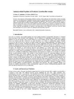
7 Refer to product labeling for further 1. Concomitant infection with A. vaginae and G. vaginalis in BV (15). Recurrence,Nugent scoreG. vaginalis detected(n = 89)G. vaginalis not detected(n = 7) vaginaenot vaginaenot detected(n = 52)(n = 37)P(n = 1)(n = 6)P BV 7-1043 (83)14 (38)< 0-69 (17)23 (62)16 Abnormal vaginal flora46 (89)19 (51)< 0-36 (11)8 (49)05 The presence of Atopobium vaginae has recently been demonstrated to serve as a useful diagnostic marker for BV (Table 1) (15-17). A study comprised of 358 women experiencing either abnormal vaginal odor or discharge evaluated the roles of Gardnerella vaginalis and Atopobium vaginae in BV recurrence following metronidazole therapy (15). Of this group, 50% were found to have normal vaginal flora, 10% were intermediate and 39% had BV as assessed by the Nugent Score criteria. Real-Time PCR analyses demonstrated the presence of Gardnerella vaginalis, often viewed as a diagnostic marker of BV, in 99% of the BV group.
8 However, 66% of the women with normal flora were also found to harbor G. vaginalis; therefore, questioning its negative predictive power and its use as a diagnostic marker (Table 2). Atopobium vaginae , however, was determined to be highly specific for BV affected individuals, with 96% detection levels observed within the BV group and only 12% within the normal flora group (Table 2) (15). Table 2. Incidence of Atopobium vaginae in BV. The presence of Atopobium vaginae serves as a better indicator of BV than does Gardnerella vaginaeGardnerella vaginalisSensitivity with relation to BV96%99%Specificity with relation to BV77%35%Prevalence associated with abnormal vaginal flora88%41%NPV96%99%PPV74%50%An inextricable link between G. vaginalis and A. vaginae has become apparent from the analyses of these BV cases in that A. vaginae is rarely ever present in the absence of G. vaginalis regardless of Nugent Score, suggesting a synergistic effect exists between the two bacteria (15-16).
9 This association was also made apparent through the analysis of vaginal biofilms, whereby G. vaginalis and A. vaginae were found to comprise greater than 90% of the film (Figure 1). The detection of high levels of both species correlates with a significant increase in antibiotic failure and the recurrence of BV, while the absence of either species is highly predictive of a negative BV diagnosis (15).Medical Diagnostic Laboratories, Thorsen P, Jensen IP, Jeune B, et al. 1998. Few microorganisms associated with bacterial vaginosis may constitute the pathologic core: a population-based microbiological study among 3,596 pregnant women. Am J Obstet Gynecol 178(3):580-587. 6. Curran D. 2006. Gardnerella. 7. Sweet RL. 1995. Role of bacterial vaginosis in pelvic inflammatory disease. Clin Infect Dis 20(Suppl 2):S271-S275. 8. Hillier SL, Nugent RP, Eschenbach DA, et al.
10 1995. Association between bacterial vaginosis and preterm delivery of low-birth-weight infant. The vaginal infections and prematurity study group. N Eng J Med 333(26):17371742. 9. Goldenberg RL, Culhane JF. 2003. Infection as a cause of preterm birth. Clin Perinatol 30(4):677-700. 10. Cohen CR, Duerr A, Pruithithada N, et al. 1995. Bacterial vaginosis and HIV seroprevalence among female commercial sex workers in Chiang Mai, Thailand. AIDS 9(9):1093-7. 11. Martin HL, Richardson BA, Nyange PM, et al. 1999. Vaginal Lactobacilli, microbial flora, and risk of human immunodeficiency virus type 1 and sexually transmitted disease acquisition. J Infect Dis 180(6):1863-8. 12. Sewankambo N, Gray RH, Wawer MJ, et al. 1997. HIV-1 infection associated with abnormal vaginal flora morphology and bacterial vaginosis. Lancet 350(9077):546-50. 13. Wiesenfeld HC, Hillier SL, Krohn MA, et al.