Transcription of Atrial Fibrillation Management of Acute Atrial ...
1 Quality Department Guidelines for Clinical Care Inpatient Atrial Fibrillation Guideline Team Team Members Jeffrey M Rohde, MD (Team Lead) Internal Medicine Sarah E Hartley, MD Internal Medicine Sarah Hanigan, PharmD Pharmacy Services Jules Lin, MD Thoracic Surgery Lewis B Morgenstern, MD Neurology (Stroke) F Jacob Seagull, PhD Medical Education David M Somand, MD Emergency Medicine David H Wesorick, MD Internal Medicine James B Froehlich MD (Consultant) Cardiology Thomas C Crawford, MD (Team Lead) Cardiology Initial Release: May, 2014 Last Reviewed: June, 2017 Inpatient Clinical Guidelines Oversight Sarah E Hartley, MD David H Wesorick, MD F Jacob Seagull, PhD For more information: 734- 936-0770 Regents of the University of Michigan These guidelines should not be construed as including all proper methods of care or excluding other acceptable methods of care reasonably directed to obtaining the same results. The ultimate judgment regarding any specific clinical procedure or treatment must be made by the physician in light of the circumstances presented by the patient.
2 Management of Acute Atrial Fibrillation and Atrial Flutter in Non-Pregnant Hospitalized Adults Patient Population. Adult hospitalized patients with Atrial Fibrillation and Flutter. This guideline excludes pregnant women. Objectives. The purpose of these inpatient care guidelines is to provide an evidence-based blue print for the Acute care of patients with Atrial Fibrillation (AF) and Atrial flutter (AFL) at the University of Michigan Health System. It is hoped that this standardization of care will result in improved patient outcomes, shorter length of hospital stay, lower readmission rates, and overall cost savings for the system. This document will discuss evaluation of patients with new onset and recurrent AF/AFL, including indications for admission, rate vs. rhythm control strategies, and plan of care following patient s discharge. Key Points Clinical Presentation Patients presenting with palpitations, irregular pulse, chest pain, dyspnea, fatigue, lightheadedness, syncope, cardio-embolic disease and new or recurrent heart failure should be evaluated for AF/AFL.
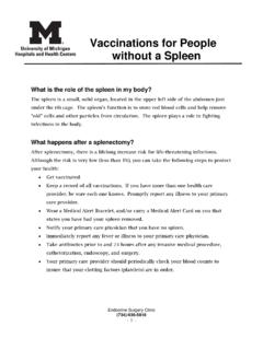
3 While AF may be asymptomatic and found incidentally, AFL is usually highly symptomatic. Diagnosis Electrocardiogram (ECG) is essential in the diagnosis of AF/AFL. The initial evaluation is summarized in Table 1 and should include: Physical exam Laboratory evaluation: CBC, basic metabolic profile, magnesium, thyroid-stimulating hormone, and cardiac enzymes as indicated Imaging: Chest X-ray, echocardiogram Continuous telemetry monitoring in the hospital Treatment Initial treatment of AF/AFL depends on hemodynamic stability: Unstable AF/AFL (Figure 1) Begin resuscitation and consider other conditions contributing to instability If instability due to AF/AFL - immediate direct current cardioversion Stable AF/AFL (Figure 2): For ED patients: Screen for early cardioversion in the Emergency Department (Figure 4) Administer rate controlling agents as indicated (Table 4) [I, B] - EP consult for uncontrolled rate despite adequate trial of rate controlling agents Consider the appropriateness of a rhythm control strategy (Table 3) [I, B] - If rhythm control strategy is appropriate/desired, consult EP and start immediate anticoagulation (Figure 3) Consider anticoagulation based on CHA2DS2-VASc score (Table 2, Figure 3) [I, A].
4 - The choice of anticoagulant will depend on the patients clinical circumstances and renal function (Figure 3) - Obtain Neurology consult prior to initiation of anticoagulation for patients with recent ischemic stroke within the prior two weeks - Patients with valvular disease and those requiring concomitant treatment with dual antiplatelet therapy should be anticoagulated with warfarin - Target-specific oral anticoagulants are preferred over warfarin in many cases * Strength of recommendation: I = generally should be performed; II = may be reasonable to perform; III = generally should not be performed. Levels of evidence reflect the best available literature in support of an intervention or test: A=randomized controlled trials; B=controlled trials, no randomization; C=observational trials; D=opinion of expert panel. 2 Figure 1: Acute Management of UNSTABLE Atrial Fibrillation and Atrial flutter (AF/AFL) ACS: Acute coronary syndrome; BNP: Brain natriuretic peptide; CBC: complete blood count; CCU: Cardiac Care Unit; COMP: comprehensive metabolic panel; CXR: chest radiograph; ECG: electrocardiogram; EP: electrophysiology; HR: heart rate; IV: intravenous; J: Joules; PE: Pulmonary embolism; RRT: rapid response team; SBP: systolic blood pressure; TSH: thyroid stimulating hormone; UA: urinalysis.
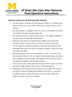
5 Figure 2: Acute Management of STABLE Atrial Fibrillation and Atrial flutter with rapid ventricular response1 3 See Figure 2 notes on following page 4 Figure 2 Notes: 1 If BP does not tolerate these medications, see Table 4 (medications for rate control) and consider DCCV or EP/General Cardiology consult. Also, IV calcium channel blockers and IV beta blockers are not usually combined- if one is not effective, change to the other. 2 If patient spontaneously converts to normal sinus rhythm: If in the ED, provide outpatient EP follow-up within 2 weeks If inpatient, consider consulting EP in the hospital If a postoperative patient, consult General Cardiology (if AF/AFL is sustained for > 24 hours) Consider a rate control agent (Table 4), depending on the pre- and post-conversion heart rate Consider the use of a 3 week event monitor after discharge to identify paroxysmal AF/AFL In all cases, consider anticoagulation (see Table 2 and Figure 3) 3 For patients with decompensated systolic heart failure: Consult cardiology, and consider digoxin or amiodarone for rate control.
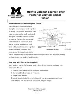
6 AV: atrioventricular; CAD: coronary artery disease; ECG: electrocardiogram; EP: electrophysiology; HR: heart rate; IV: intravenous; LV: left ventricular; WPW: Wolff Parkinson White. 5 Figure 3: Management of anticoagulation therapy in Atrial Fibrillation and Atrial flutter1 6 Figure 4: Emergency Department screening for early cardioversion of Atrial Fibrillation and Atrial flutter CrCl: creatinine clearance; SBP: Systolic blood pressure. 7 Table 1: Diagnostic evaluation of Atrial Fibrillation and Atrial flutter Current electrocardiogram (ECG) Complete physical exam Current Basic Metabolic Panel, Magnesium, complete blood count (CBC) Current thyroid stimulating hormone (TSH) Chest X-ray (CXR) Serial cardiac enzymes (Troponin, CK, CK-MB) as indicated Continuous telemetry monitoring Echo within the past 6 months to assess for the presence and severity of structural heart disease Table 2: CHA2DS2 VASc score and annual stroke risk C Congestive heart failure (or Left ventricular systolic dysfunction) 1 H Hypertension.
7 Blood pressure consistently above 140/90 mmHg (or treated hypertension on medication) 1 A2 Age 75 years 2 D Diabetes Mellitus 1 S2 Prior Stroke or TIA or thromboembolism 2 V Vascular disease ( peripheral artery disease, myocardial infarction, aortic plaque) 1 A Age 65 74 years 1 Sc Sex category ( female gender) 1 Table 3: When to consider a rhythm control strategy for Atrial Fibrillation /flutter First occurrence of symptomatic AF/AFL Occurrence or recurrence of AF due to reversible cause ( hyperthyroidism, pulmonary embolism, postoperative state, pneumonia, Acute coronary syndrome/ Acute myocardial infarction ACS/AMI) Hospital readmissions for AF/AFL or Management of AF-related comorbidities Atrial Tachyarrhythmia-related symptoms despite adequate rate control, or inability to achieve adequate rate control Cardiomyopathy presumed to be secondary to tachycardia Younger patients (age < 65), even with minimally symptomatic or asymptomatic AF/AFL CHA2DS2-VASc Score Stroke Risk (%/year) 0 0 1 2 3 4 5 6 7 8 9 8 Table 4: Pharmacologic agents useful for rate control in patients with AF/AFL Drug When to consider use?
8 [level of recommendation / level of evidence] Warnings/ Contraindications Intravenous Dosing Oral dosing/ notes Calcium Channel Blockers Diltiazem See Figure 2 [Class I, LOE B] Can cause hypotension and AV nodal block. CI: Bradycardia CI: Systolic heart failure IV: 10-20 mg IVP bolus over 2 min. If HR remains > 120, consider a second bolus over 2 minutes Then start infusion at 5 mg/hr, titrate by mg/hr every 30 minutes to HR, maximum dose 15 mg/hr [onset time 2-7 min] 120 to 360 mg daily in divided doses; extended- release preparation is available [onset 2 to 4 hours] Verapamil Can be used as an alternative to diltiazem. [Class I, LOE B] Can cause hypotension and AV nodal block. CI: Bradycardia. CI: Systolic heart failure IV: mg IVP over 2 minutes; second dose of 5-10 mg (~ mg/kg) may be given 15-30 minutes after the initial dose if patient tolerates, but does not respond to initial dose; maximum total dose: 20-30 mg. [onset time 3-5 min] 120 to 360 mg daily in divided doses; extended-release preparation is available [onset 1 to 3 hours] Beta-blockers Metoprolol See Figure 2 [Class I, LOE C] Can cause hypotension and AV nodal block.
9 Avoid in patients with bronchoconstriction or emphysema. Use with caution in patients with decompensated heart failure. CI: Bradycardia. IV: 5 mg IVP every 10-20 minutes x 3 [onset time 5 min]. If HR < 120 results, start oral metoprolol (see dosing suggestions to right). If HR > 120 after 15 mg IV metoprolol, consider an alternate agent such as diltiazem above. Oral dosing: If HR < 120 after 5 mg IV, consider oral dose of 25 mg PO BID. If HR < 120 after 10 mg IV, consider oral dose of 50 mg PO BID. If HR < 120 after 15 mg IV, consider oral dose of 75 mg PO BID. [onset 4-6 hours] Esmolol Best for use in situations where there is an indication to use a beta-blocker, but the patient a.) cannot take oral medications, or b.) has a labile or tenuous blood pressure (esmolol has a short half-life, and its effect can be quickly halted by stopping the infusion). [Class I, LOE C] Can cause hypotension and AV nodal block. Avoid in patients with Acute or active airway obstruction/ bronchoconstriction.
10 Avoid in patients with decompensated heart failure. Relatively expensive CI: Bradycardia. IV: 50 mcg/kg/minute infusion for 4 minutes. Infusion may be continued at 50 mcg/kg/minute or, if the response is inadequate, titrated upward in 50 mcg/kg/minute increments (increased no more frequently than every 4 minutes) to a maximum of 200 mcg/kg/minute. [onset time < 5 min] N/A Loading doses are not necessary due to rapid onset of action Propranolol Consider for use in thyrotoxicosis [Class I, LOE C] Can cause hypotension and AV nodal block. Avoid in patients with Acute or chronic airway obstruction/ bronchoconstriction. Avoid in patients with decompensated heart failure. CI: Bradycardia. IV: mg over 1 minute; may repeat, if necessary, up to a total maximum dose of mg/kg [onset time 5 min] Usual oral dose: 10-30 mg/dose every 6-8 hours 9 Table 4: Pharmacologic agents useful for rate control in patients with AF/AFL, continued Drug When to consider use?