Transcription of BD Cell Viability Kit
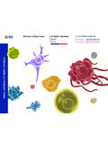
1 Live and Dead .. BD Cell Viability Kit Cell Catalog No. Number of tests Discrimination 349483 100 Tests 349480 with BD Liquid Counting Beads 100 Tests .. RESEARCH Studies of: APPLICATIONS. rapid counting of live/dead bacteria or other microbial cells1-4. efficacy of bacterial disinfectant5. Viability of yeast during fermentation6. Viability and concentration of cells in culture Viability and concentration of cells before staining for flow cytometric analysis mammalian or microbial cells Viability and count of cells in bioreactors Viability of sperm for research studies7,8. Viability in cell preparations containing debris DESCRIPTION Flow cytometry provides a rapid and reliable method to quantify viable cells in eukaryotic and prokaryotic cell ,7,8 The BD Cell Viability Kit offers an easy-to-use dye combination to distinguish live and dead cells for analysis by flow cytometry.
2 The kit contains thiazole orange (TO) solution to stain all cells and propidium iodide (PI) to stain dead cells . BD Liquid Counting Beads is a liquid suspension of fluorescent beads. Add the beads to a flow sample to calculate absolute counts. The kit can be ordered with or without counting beads. The method provides a rapid alternative to manual microscopic Live cells have intact membranes and are impermeable to dyes such as PI, which leaks into cells with compromised membranes. TO is a permeant dye and enters all cells , live and dead, to varying degrees. The fluorescent signal from TO in viable cells allows their enumeration even when debris in the cell preparation contaminates a scatter gate around the cells . Thus the combination of these two dyes provides a rapid and reliable method for discriminating live and dead eukaryotic and prokaryotic cells , including peripheral blood mononuclear cells (PBMCs), mammalian cell lines, bacteria, and yeast.
3 MATERIALS PROVIDED The kit contains 1 vial of 500 L 42 mol/L TO in dimethyl sulfoxide (DMSO) and 1 vial of 500 L mmol/L PI in water. The optional BD Liquid Counting Beads are supplied as 1 vial of 10 mL of fluorescent microspheres in buffer with sodium azide. Material required but not Recommended staining buffer: Physiologic phosphate-buffered saline containing provided Pluronic F68 and 1 mmol/L EDTA. For Research Use Only. Not for use in diagnostic or therapeutic procedures. Becton, Dickinson and Company BD Biosciences 2350 Qume Drive San Jose, CA 95131 USA. 5/2015 23-6755-03. NOTE For bacteria, TWEEN 20 can be substituted for the Pluronic F68. The staining buffer should be passed through a m filter. HANDLING AND Store vials at 2 8 C with the TO and PI stored in the desiccated container provided. STORAGE Each reagent is stable for the period shown on the bottle label when stored as directed.
4 WARNINGS All biological specimens and materials coming in contact with them are considered biohazards. Handle as if capable of transmitting infection9,10 and dispose of with proper precautions in accordance with federal, state, and local regulations. Never pipette by mouth. Wear suitable protective clothing, eyewear, and gloves. METHOD Recommended amounts of TO and PI depend on the cell type being stained. Dilute cultured cells or PBMCs at least 1:10 in staining buffer to an approximate concentration range of 5 x 10⁵ to 107 cells /mL. (See Material required but not provided for preparation method.) For other sample types, such as pharmaceutical, food or environmental samples, at least 100 organisms per mL need to be detected using flow cytometry. If necessary, samples can be brought into this range by an initial concentration step.
5 Bacteria and yeast: Add L of each dye solution to 500 L of cell suspension. The final staining concentrations are 420 nmol/L for TO and 43 mol/L for PI. Vortex and incubate for at least 5 minutes at room temperature. Mammalian cells : Add L of TO and L of PI solution to 2 mL of cell suspension. The final staining concentrations are 84 nmol/L for TO and mol/L for PI. Vortex and incubate for 5 minutes at room temperature. Bull semen: Add L of each dye solution to 2 mL of cell suspension. The final staining concentrations are 42 nmol/L for TO and mol/L for PI. Vortex and incubate for 5 minutes at room temperature. NOTE When using BD Liquid Counting Beads, allow the beads to come to room temperature. Prior to analysis, gently vortex the bead suspension for 30 seconds and add 50 L to each tube. Use the reverse pipetting technique to pipette for better accuracy.
6 Cap the tubes and gently vortex to mix. Acquisition and analysis Acquire prepared samples on a BD FACS brand flow cytometer equipped with 488-nm laser excitation and BD CellQuest software. Use an FSC threshold for mammalian cells (fluorescence threshold if also using BD Liquid Counting Beads), and an SSC threshold for microbial cells . Gate cells using scatter and TO fluoresces primarily in FL1 and FL2; PI fluoresces primarily in FL3. Therefore, the best discrimination of live and dead populations is on an FL1 vs FL3 plot. To determine the concentration of the cell populations, use the following equation: # events in cell region- # beads/test*. ---------------------------------------- ------------------ --------------------------------- dilution factor = concentration of cell population # events in bead region test volume * This value is found on the vial of BD Liquid Counting Beads and can vary from lot to lot.
7 Representative data Figure 1 shows results obtained by adding 50 L of BD Liquid Counting Beads to a 500- L sample of E. coli stained with 5 L of TO and 5 L of PI. Regions are set around the live, injured, and dead bacterial populations; counting beads are not shown. 23-6755-03 Page 2. Figure 1 FL1 vs FL3 dot plot, gated on E. coli by scatter R6. dead R5. injured FL3-H. R4. live FL1-H. Figure 2 shows results obtained by adding 50 L of BD Liquid Counting Beads to a 2-mL sample of PBMCs stained with 4 L of TO and 2 L of PI. Live cells are in R5;. dead cells are in R3; and counting beads are in R4. Figure 2 FL1 vs FL3 dot plot, gated on PBMCs by scatter R3. dead beads R4. FL3-H. injured R5. live FL1-H. Figure 3 shows results obtained by adding 50 L of BD Liquid Counting Beads to a 2-mL sample of Raji cells stained with 4 L of TO and 2 L of PI.
8 Live cells are in R5;. dead cells are in R3; and counting beads are in R4. Figure 3 FL1 vs FL3 dot plot, gated on Raji cells by scatter R3. dead beads R4. FL3-H. injured live R5. FL1-H. Figure 4 shows results obtained by staining a 2-mL sample of thawed bull semen with 2. L of TO and 2 L of PI. For staining, 20 L of thawed semen was added to 2 mL of staining buffer. Live sperm are in R3, dead sperm are in R2, and injured sperm are between the two regions. Page 3 23-6755-03. Figure 4 FL1 vs FL3 dot plot, gated on thawed bull semen by scatter R2. dead FL3-H. injured R3. live FL1-H. LIMITATIONS Dispose of stained samples, extra dye solution, and container according to local, state, and federal regulations. CHARACTERIZATION To ensure consistently high-quality reagents, each lot of reagent is tested for conformance with characteristics of a standard reagent.
9 Representative flow cytometric data is included in this data sheet. WARRANTY Unless otherwise indicated in any applicable BD general conditions of sale for non-US. customers, the following warranty applies to the purchase of these products. THE PRODUCTS SOLD HEREUNDER ARE WARRANTED ONLY TO CONFORM TO THE QUANTITY AND CONTENTS. STATED ON THE LABEL OR IN THE PRODUCT LABELING AT THE TIME OF DELIVERY TO THE CUSTOMER. BD. DISCLAIMS HEREBY ALL OTHER WARRANTIES, EXPRESSED OR IMPLIED, INCLUDING WARRANTIES OF. MERCHANTABILITY AND FITNESS FOR ANY PARTICULAR PURPOSE AND NONINFRINGEMENT. BD'S SOLE LIABILITY. IS LIMITED TO EITHER REPLACEMENT OF THE PRODUCTS OR REFUND OF THE PURCHASE PRICE. BD IS NOT LIABLE. FOR PROPERTY DAMAGE OR ANY INCIDENTAL OR CONSEQUENTIAL DAMAGES, INCLUDING PERSONAL INJURY, OR ECONOMIC LOSS, CAUSED BY THE PRODUCT.
10 REFERENCES 1. Nebe-von-Caron G, Stephens PJ, Badley AR. Bacterial detection and differentiation by cytometry and fluorescent probes. Proc Royal Microbiol Society. 1999;34:321-327. 2. Nebe-von-Caron G, Stephens PJ, Hewitt CJ, Powell JR, Badley RA. Analysis of bacterial function by multi-colour fluorescence flow cytometry and single cell sorting. J Microbiol Meth. 2000;42:97-114. 3. Davey HM, Kell DB. Flow cytometry and cell sorting of heterogeneous microbial populations: the importance of single-cell analyses. Microbiol Rev. 1996;60:641-696. 4. Alsharif R, Godfrey W. Bacterial detection and live/dead discrimination by flow cytometry. Microbial Cytometry Application Note. BD Biosciences, Immunocytometry Systems; San Jose, CA. 2002. 5. Alsharif R, Tapia M, Godfrey W, Wanalund J, Nagar M. Bacterial disinfectant efficacy using flow cytometry.