Transcription of Benign Paroxysmal Positional Vertigo
1 Benign Paroxysmal Positional VertigoDavid Solomon, MD, PhDAddressDepartment of Neurology, University of Pennsylvania, 3 W. Gates Building, 3400 Spruce Street, Philadelphia, PA 19104-4283, : Treatment Options in Neurology 2000, 2:417 427 Current Science Inc. ISSN 1092-8480 Copyright 2000 by Current Science Paroxysmal Positional Vertigo (BPPV) is the mostcommon diagnosis made in many specialty clinics serv-ing patients with dizziness. This diagnosis is suggested bya history of brief (less than one minute) episodes of ver-tigo that are provoked by rolling over in bed, lying down,sitting up from a supine position, bending over, or look-ing up. BPPV commonly is worse in the early morning(matutinal Vertigo ), and may be absent for weeks ormonths at a time before returning. Diagnosis rests on theobservation of characteristic eye movements accompany-ing the symptoms of Vertigo when a patient s head ismoved into a specific orientation with respect to and Hallpike provided both the provocative maneu-ver necessary for the accurate diagnosis of the condition,as well as the first description of all the classic features ofthe accompanying nystagmus: latency, direction, dura-tion, reversal, and fatigability [2 ].
2 BPPV occurs when free particles, suspended in thefluid (endolymph) of a patient s vestibular labyrinth, findtheir way into one of the semicircular canals (SCC). Nor-mally, the canals are excited only by head rotation; whenparticles more dense than endolymph are present in thelumen, however, canals become gravity sensitive, patho-logically responding to changes in head position [3,4]. Atleast in some cases, the particles (canaliths) are otoconia calcium carbonate crystals, a normal constituent of theotolith organs in the inner ear [5]. Original theories heldthat the particles were adherent to the cupula, the struc-ture that spans the lumen of the canal and is deflectedduring head rotation [6]. More recently, free-floatingmaterial has been observed intraoperatively in the poste-rior SCC [7].
3 Convincing evidence [8, Class I] in supportof canalithiasis as a pathophysiologic explanation forBPPV validates treatment with any procedure that caneffectively clear these dense particles from the posteriorsemicircular canal. Canalithiasis can occur in any posterior semicircular canal (PSC) was affected in themajority of cases of BPPV (93% of cases) [9], with 85%being unilateral, and 8% affecting the PSC on both horizontal semicircular canal (HSC) was affected in5% of cases. Involvement of an anterior canal is of the features of typical nystagmus provoked bychanges in head position in patients with BPPV may beexplained by canalithiasis [4]. Latency is typically on theorder of a few seconds, although cases with greater than10 second latency have been described. Because eachcanal has excitatory connections to extraocular musclesthat move the eyes in the same plane (Fig.)
4 1), the direc-tion of the nystagmus depends on the canal that is beingstimulated (Tables 1 and 2), and the direction of parti-cle movement. The nystagmus seen when lying downmay reverse direction when the patient moves to theupright position. The duration of nystagmus cor-responds with the time needed for particles to come torest in a new dependent location. Although fatigabilityis described as a characteristic of nystagmus associatedwith BPPV, repeated positioning is not recommended,as the diagnosis may be made without subjecting thepatient to additional discomfort. Also, there is goodreason to expect that procedures used to clear particlesfrom the canal may be less efficacious if the debris isdispersed by repeatedly being moved in the statementBenign Paroxysmal Positional Vertigo can be diagnosed with great certainty, and treated effectively at the bedside using one of the canalith repositioning procedures described in this paper.
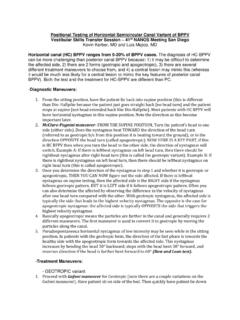
5 This treatment has been shown effective in properly controlled trials, has a rational basis, and has minimal risk [1].418 Neurologic Ophthalmology and OtologyFigure this view of vestibular and orbital anatomy from above, the dotted lines represent the planes containing the poste-rior semicircular canal (PSC) of the right labyrinth, the superior and inferior recti of the left eye, and the superior and inferior oblique muscles of the right eye. This corre-sponds to the main neuroanatomical con-nections of the vestibular ocular reflex. Activation of the PSC, therefore, results in a mixed vertical and torsional nystagmus, with the contralateral eye having more upbeat, and the ipsilateral eye more extorsional 1. Direction, latency, and duration of observed nystagmus IpsilateralContralateralQuick phaseInferior oblique (extorsional)Superior rectus (upward)Slow phaseSuperior oblique (intorsional)Inferior rectus (downward)Table 2.
6 Positional tests for horizontal canal BPPVHead postionNystagmus directionAffected sideMechanismRight ear downGeotropic (R beat)Stronger nystagmus with affected ear downCanalithiasisLeft ear downGeotropic (L beat)CanalithiasisRight ear downAgeotriopic (L beat)Stronger nystagmus with affected ear upCupuloithiasisLeft ear downAgeotriopic (R beat)CupuloithiasisBenign Paroxysmal Positional VertigoSolomon419 Treatment Predisposing factors of BPPV: Circumstances in which the head is placed or maintained in an inverted orientation (eg, dental procedures, visits to the hairdresser). Age. Inactivity. Trauma and vestibular neuritis. Other ear disease; Meniere s syndrome [10]. Family history [11].Antiemetic therapyPromethazineStandard dosagePromethazine is administered orally at mg, 25 mg, or 50 mg prior to treat-ment, or as a suppository at the same doses, if the patient is already symptomatic with vomiting.
7 May be repeated every 4 to 6 not be used in those patients with adverse reactions to phenothiazines. Use with caution in those patients with narrow-angle glaucoma, and obstructive bladder drug interactionsMay increase sedative action of other central nervous system depressants (alcohol, narcotics, and so forth).Main side effectsSedation, and dry mouth. Rarely, extrapyramidal motor manifestations (oculogyric crisis, dystonic reaction) pointsVestibular suppressants do not affect the Vertigo associated with this condition [12]. At most, they may reduce the motion sickness, which frequently accompanies the attacks. Patients are routinely requested to stop symptomatic therapy prior to electronystagmogram (ENG) testing or examination, because a gaze evoked nystag-mus, or pursuit abnormalities, may be caused by some vestibular suppressants.
8 In general, there are no indications for the chronic use of vestibular suppressants such as meclizine. These may be used on an as needed basis to treat the motion sickness symptoms associated with attacks of labyrinthine Vertigo , but habitual use will retard the process of central nervous system compensation following any change in peripheral vestibular effectivenessOnly few doses should be required, and this medication is available in an inexpen-sive, generic form. Use of a canalith repositioning procedures (CRPs), such as the Epley and Semont maneuvers, depends on the accurate localization of particles. Examining physicians must determine the ear that is affected, which canal in the identified ear is affected, and whether the material is free-floating or adherent to the test for posterior semicircular canal BPPVD iagnosis of BPPV affecting the PSC is made by observing the typical upbeat and torsional nystagmus (Table 1, Fig.)
9 1) after performing the Dix-Hallpike maneuver. This is shown in the first two panels of Figure 2. The patient is seated with legs extended on the examining table, and the head is turned 45 to either the left or right. The patient is then brought into the supine position briskly, but this need not be done too rapidly. After observing for eye movements and questioning the Pharmacologic treatmentInterventional procedures420 Neurologic Ophthalmology and Otologypatient about symptoms, the patient is brought back to the sitting position. The head should be maintained in the same orientation with respect to the body during the entire maneuver. The fellow ear is then tested by rotating the head in the other direction and repeating the positioning. The patient must be brought upright between each ear , fixation is removed by monitoring eye movements using either an infrared camera system in darkness, or by using Frenzel lenses.
10 Electro-oculography is insensitive to torsional eye movements, and two-dimensional video-oculography likewise may give spurious results. Even without removal of fixation, the torsional component of the nystagmus is not suppressed, and should remain observable. In making the diagnosis of BPPV, direct observation is therefore more helpful than laboratory upbeat-torsional nystagmus will be generated by the vestibular ocular reflex (VOR) as the patient is being moved, and the patient will correctly perceive head rotation. When the head comes to rest in its new orientation, normally, there is no further nystagmus or sense of movement. In the patient with canalithiasis (parti-cles in the duct of the SSC), a second excitation occurs as the debris gravitates to a new lowest point. This induces a response by the canal indistinguishable from that caused by actual rotation of the head in space there is the same nystagmus and associated perception of rotation, now termed Vertigo , which persists for as long as the particles are in the quick phases of nystagmus are noted clinically, the actions of the superior rectus and inferior oblique account for the up beating and torsional direc-tions, respectively (Table 1).