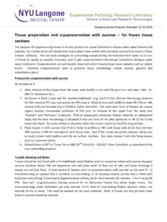
Transcription of Bone mineral density (BMD) and tissue mineral density (TMD ...
1 HU and BMD calibration in Bruker-MicroCT CT-analyser Page 1 of 30 Method note Bone mineral density (BMD) and tissue mineral density (TMD) calibration and measurement by micro-CT using Bruker-MicroCT CT-Analyser HU and BMD calibration in Bruker-MicroCT CT-analyser Page 2 of 30 1. INTRODUCTION What is density measured by x-ray micro-CT? A micro-CT 3D image of an object is basically a 3D map of local x-ray absorption in the object. The reconstruction from multiple angular views provides information on how much x-ray absorption is happening within each cubic voxel element of the scanned volume.
2 It is important to understand what is meant by density in a microCT image. The reconstructed grey-scale intensity of each image voxel does not relate directly to mass density alone, thus do not expect it to correspond with measurements of weight per volume, The primary entity that is measured is x-ray absorption, defined as the attenuation coefficient in units of 1/distance, specifically, mm-1. This is determined both by mass density and elemental composition of the material. However, when we know that the x-ray absorption of a material is dominated by one specific material, then we can relate the measured x-ray attenuation coefficient (AC) to the mass density of that material.
3 The calibration of bone mineral density (BMD) is an example of this. We assume that the x-ray attenuation within mineralised tissues such as bone, dentine and enamel is dominated by, and can be approximated as, the x-ray attenuation of the mineral compound calcium hydroxyapatite (CaHA) which has the formula Ca5(PO4)3(OH). Then we obtain phantoms with two or more known mass concentrations of CaHA. By linking these mass concentrations of CaHA with measured x-ray AC in the microCT image, we can establish calibration linking the two quantities, allowing us to infer from the microCT measured attenuation coefficient in a mineralised tissue , the density of CaHA in.
4 Note that this calibration system could be used to calibrate measurement of other substances apart from CaHA and bone, where x-ray absorption is known to be dominated by a certain substance and phantoms can be prepared with known concentrations of that substance. Definitions of BMD and TMD Bone mineral density (BMD) is defined as the volumetric density of calcium hydroxyapatite (CaHA) in a biological tissue in terms of It is calibrated by means of phantoms with known density of CaHA. Bone mineral density can refer to two different measurements: HU and BMD calibration in Bruker-MicroCT CT-analyser Page 3 of 30 (a) the combined density of a well-defined volume which contains a mixture of both bone and soft tissue , such as a selected volume of medullary trabecular bone in a femur or tibia, is measured as bone mineral density , or BMD.
5 This parameter BMD relates to the amount of bone within a mixed bone-soft tissue region, but does not give information about the material density of the bone itself. (b) the density measurement restricted to within the volume of calcified bone tissue , such as cortical bone, excluding surrounding soft tissue , is called tissue mineral density or TMD [1]. By contrast to BMD, this gives us information about the material density of the bone itself, and ignores surrounding soft tissue . Typically trabecular bone is assessed for BMD, averaging bone and marrow within the medullary volume of interest.
6 By contrast, for cortical bone, TMD is measured. Note that in the case of trabecular bone, partial volume effect compromises the measurement of TMD except at very high micro-CT resolutions. As a general rule, resolution should be such that the thickness of a bone structure should equal 10 pixels or more, in order for TMD to be measured. Bruker-MicroCT BMD/TMD calibration phantoms: size is important! Bruker-MicroCT BMD calibration phantoms are in pairs, with concentrations of CaHA of and Due to beam hardening effects, size of an object slightly affects measured density , as shown in figure 1 for rods of CaHA (in epoxy resin) at thicknesses of 2-16 mm.
7 Figure 1. Increasing diameter rods of the same material (with CaHA) show a small decline in measured attenuation coefficient, due to beam hardening. HU and BMD calibration in Bruker-MicroCT CT-analyser Page 4 of 30 This means that it is important to use a calibration rod which approximately matches the crossectional area of calcified tissue of the bone or tissue sample within which you wish to measure bone mineral density . Figure 2. Bruker-MicroCT BMD calibration rod pairs are provided in a range of diameters (2-4mm, 8-16mm, 32mm) to match the size of the scanned bone.
8 Bruker-MicroCT provides calibration rod pairs, composed of epoxy resin with embedded fine CaHA powder at concentrations of and , and at diameters of 2mm, 4mm, 8mm, 16mm and 32mm. The size of phantom to be used for different bones is suggested in table 1 below. Table 1. The five diameters of Bruker-MicroCT BMD calibration rod pairs are recommended for bones of corresponding size. BMD phantom rod pair diameter Appropriate bone types 2mm Mouse bones (femur, tibia, vertebra etc.) 4mm Rat bones (femur, tibia, vertebra etc.) 8mm 8mm trephine biopsy cores, rabbit bones, human teeth, bones of primates, dogs 16mm Larger bones such as sheep bones 32mm Bones above 3cm in diameter, such as human bones, bovine, etc.
9 HU and BMD calibration in Bruker-MicroCT CT-analyser Page 5 of 30 Simulate surrounding material around bone in calibration scans Not only does the thickness of a scanned material such as bone affect its measured density . Soft tissue such as muscle, fat and skin around a bone will also significantly affect the measured density of the bone. Therefore, if rodent limbs are scanned in vivo, the corresponding BMD calibration phantoms should be scanned in a tube of water ( eppendorf) approximately matching the width of the animal s leg (figure 3). Water closely imitates the x-ray absorption of soft biological tissue in general.
10 (a) (b) Figure 3. An in vivo scan of a rodent limb (a) should be calibrated for BMD measurement by an equivalent scan of the BMD rod pair of appropriate size (mouse 2mm, rat 4mm) with the rods scanned inside a tube of water (b), the tube matching approximately the animal s leg diameter. This is to simulate x-ray absorption of the surrounding tissue , for improved accuracy of BMD measurement. The water tube diameter for example should be about 6-8 mm for the mouse knee, 10-15 mm for the rat knee. Note that it is not too important for the BMD rods to be in the center of the water tube they can lie against the side, the effect of the water on x-ray absorption averaged over 180 or 360 degrees of scanning will be similar.
![transition to retirement TUSM-FINALrev02-17-10[1]](/cache/preview/2/0/8/1/d/3/1/7/thumb-2081d317aceb3fadbe14f98091ebcdcd.jpg)