Transcription of Breast Development - Columbia University
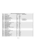
1 1 Breast PathologyLecturer: Hanina Hibshoosh, : Kumar, Cotran, Robbins, Basic Pathology, 6th Edition, pages 623 - 635 Breast Development 5th week - thickening of the epidermis - milk line formation Mammary ridges form from axilla to groin region Involution of mammary ridges except in chest region, persistance yield supernumerary Breast (polythelia) 15th week - downward growth into stroma Last two months of gestation canalization of epithelial cords with formation of branching and lobuloalveolarstructures234 Fibrocystic Changes Miscellaneous changes of Breast involving ducts, lobules, and stroma Clinical incidence approximately 40 to 50% of patients, lumps Pathological incidence greater than 60 to 80% Terminology - fibrocystic change favored over disease Pathogenesis - reflects exaggerated changes occurring normally in the menstrual cycle5 Fibrocystic Disease of Breast1. Fibrosis2. Cysts3. Apocrine Metaplasia4. Sclerosing Adenosis5. Papillomatosis (epithelial hyperplasia)678910 Consensus Statement Cancer Committee of the College of American PathologistsRelative Risk for Invasive Breast Cancer Following Pathological Diagnosis of Benign Breast DiseaseNo increased riskSlightly increased risk ( )Hyperplasia, mild (2-4 cells)Hyperplasia, moderate or floridApocrine metaplasia (ductual and lobular)Cysts (macro and micro) Papilloma with fibrovascular coreDuct ectasiaAdenosis (sclerosing or florid)FibroadenomaModerately increased risk (5x)Atypical Ductal and Lobular Hyperplasia-Borderline Lesions11 Atypical Ductal Hyperplasia (ADH)Borderline lesion is defined by some as a proliferative lesion in which some of the cytologic and architectural criteria of carcinoma in situ are met but are not fully satisfied.
2 Page and Rogers have more recently published criteria including qualitative and quantitative features in the classification of ductal and lobular proliferative ) Cytologic criteria- This guideline emphasizes the need for a population of cells bearing similarity to neoplastic ) Histologic or architectural criteria- the ductal epithelial proliferation predominant pattern is cribiform with secondary spaces having both punched-out regular (typical of non-comedo carcinoma in situ) and irregular borders (typical of florid hyperplasia without atypia).c) Quantitative criteria - anatomic extent of lesion. At least two basement membrane-bound spaces need to be completely involved by a cell populationwith the cytologic and histologic features of ductal carcinoma in situ (DCIS) for a diagnosis of DCIS. Therefore, a lesion with a single space with diagnostic features of DCIS is diagnosed as atypical ductal hyperplasia. In addition, support for an overall size criterion such as advocated by Tavassoli (2mm cumulative changes) is Incidence approximately 5% Risk for developing invasive cancer4-5x general population Approximately 10% of patients will develop cancer Risk for cancer is bilateral Risk greatest in first decade following diagnosis Patients with atypical ductal hyperplasia with family history have same risk as in-situ cancer group for the Development of invasive cancer, 8-10x.
3 Prognosis of atypical ductalhyperplasia associated cancer same as cancer lacking atypical ductal hyperplasia ~30% of patients with ADH on needle biopsy have cancer on excisionAtypical Ductal Hyperplasia (ADH)13 Benign Neoplasma Fibroadenoma Intraductal papillomaTumor of variable malignant potential Cystosarcoma phyllodesFibroadenoma Most common benign tumor of the female Breast Usually appears in young women Peak incidence in the third decade of life A benign fibroepithelial tumor usually solitary may be multiple Rarely associated with carcinoma141516 Intraductal Papilloma A benign papillary neoplasm within a duct Identified peripherally or centrally (nipple duct) in which case it may be associated with bloody nipple discharge Mild increased risk ( - 2x) of Development of invasive cancer in patients with multiple peripheral intraductal papillomas1718 Cystosarcoma Phyllodes A fibroepithelial neoplasm of variable malignant potential Neoplastic component is the stroma Degenerates into a frank sarcoma with metastasis to lung and distant organs The majority can be cured by complete excision Peak incidence at 50 years of age192021 Malignant Breast Lesions Epithelial derived tumors Intraductal and invasive ductal carcinoma In situ and invasive lobular carcinoma Mesenchymal neoplasm (sarcoma) Cystosarcoma phyllodes Angiosarcoma OthersPathology of Breast Carcinoma Most cancers (90%) show ductal epithelium differentiation 10% referred to as lobular type In situ and invasive components22 Ductal Carcinomas In Situ Neoplastic transformation of ductal epithelium within ducts or lobules (intraductal) confined by basement membrane Various histologic patterns.
4 Comedo, solid, cribiform, clinging, and papillary type May be detected by its association with microcalcifications May represent up to 25% of Breast carcinoma High grade and large size of the in situ carcinoma predicts multifocality and propensity for invasion Relative risk for the Development of an invasive carcinoma 8-10 fold greater than the general population Risk is primarily ipsilateral232425 Paget s Disease of the Breast In situ carcinoma of lactiferous ducts with extension to epidermis Involving the nipple and areola May present with nipple discharge, crusting, or excoriation of nipple surface2627 Invasive Ductal Carcinoma An infiltrative malignant epithelial process resembling cells lining ducts--most common Breast carcinoma Classified according to histologic appearance as:1. Carcinoma not otherwise specified -- majority2. Special good prognosis subtype including medullary carcinoma, colloid (mucinous) carcinoma, and tubular carcinoma.
5 3. Poor prognosis -- inflammatory carcinoma 28293031 Lobular Carcinoma In Situ (LCIS) Neoplastic transformation of epithelial cells lining terminal ducts and acini of small size E-cadherinnegative Typically multifocal and bilateral 6 - 9 fold increased risk for Development of invasive cancer Bilateral risk for Development of invasive cancer 3/4 of invasive cancer are of ductal type Considered primarily a marker for invasion32 Invasive Lobular Carcinoma An infiltrating carcinoma resembling cells lining the lobules (of LCIS) Histologically showing classical Indian file pattern and targetoid bull s eye pattern Composed of relatively small cells with scanty cytoplasm, sometimes vacuolated (E-cadherinnegative) Represents approximately 10% of Breast cancer with a higher than usual incidence of bilaterality(approximately 20%)33 Prognostic Factors of Breast Cancer Size of primary tumor Lymph node involvement and extent Grade Histologic type To a lesser extent, estrogen/progesterone receptors, S-phase fraction but not DNA content, C-Erb B2 and.
6 34 Gynecomastia Button-like subareolar swelling, bilateral, histologically corresponding to intraductal epithelial hyperplasia and increased periductal stromalcellularity and edema Associated with relative estrogen excess, cirrhosis of liver, Klinefelter s, estrogen secreting tumor, estrogen therapy, and digitalis therapy Physiological gynecomastia most common in puberty and old age No clear cut association with Development of carcinoma35 Male Breast Carcinoma Rare, ratio of male to female Breast cancer 1:125 Occurs in advanced age Identified in peri-nipple/areolar region Presents in advanced stage Resembles morphologically and biologically invasive carcinomas of the female Breast