Transcription of BTS guidelines for the insertion of a chest drain
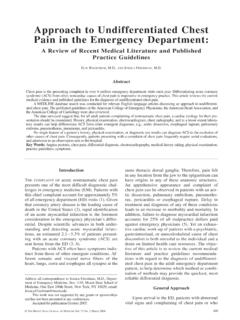
1 bts guidelines for the insertion of a chest drainD Laws, E Neville, J Duffy, on behalf of the British Thoracic Society Pleural DiseaseGroup, a subgroup of the British Thoracic Society Standards of Care ;58(Suppl II):ii53 ii591 BACKGROUNDIn current hospital practice chest drains are usedin many different clinical settings and doctors inmost specialities need to be capable of their safeinsertion. The emergency insertion of a large borechest drain for tension pneumothorax followingtrauma has been well described by the AdvancedTrauma and Life Support (ATLS) recommenda-tions in their instructor s manual1and there havebeen many general descriptions of the step bystep method of chest tube 9It has been shown that physicians trained in themethod can safely perform tube thoracostomywith 3% early complications and 8% theseguidelines we discuss the safe insertion of chesttubes in the controlled circumstances usuallyencountered by physicians. A summary of theprocess of chest drain insertion is shown in fig TRAINING All personnel involved with insertion ofchest drains should be adequately trainedand supervised.
2 [C]Before insertion of a chest drain , all operatorsshould have been adequately trained and havecompleted this training appropriately. In all othercircumstances, insertion should be supervised byan appropriate is part of the SHO corecurriculum training process issued by the RoyalCollege of Physicians and trainees should beexpected to describe the indications and compli-cations. Trainees should ensure each procedure isdocumented in their log book and signed by thetrainer. With adequate instruction, the risk ofcomplications and patient pain and anxiety canbe guidelines will aid the training of juniordoctors in the procedure and should be readilyavailable for consultation by all doctors likely tobe required to carry out a chest tube INDICATIONSC hest tubes may be useful in many settings, someof which are listed in box PRE-DRAINAGE RISK ASSESSMENT Risk of haemorrhage: where possible, anycoagulopathy or platelet defect should becorrected prior to chest drain insertionbut routine measurement of the plateletcount and prothrombin time are only rec-ommended in patients with known riskfactors.
3 [C] The differential diagnosis between apneumothorax and bullous disease re-quires careful radiological it is important to differentiatebetween the presence of collapse and apleural effusion when the chest radio-graph shows a unilateral whiteout . Lung densely adherent to the chest wallthroughout the hemithorax is an absolutecontraindication to chest drain insertion .[C] The drainage of a post pneumonectomyspace should only be carried out by orafter consultation with a cardiothoracicsurgeon. [C]There is no published evidence that abnormalblood clotting or platelet counts affect bleedingcomplications of chest drain insertion . However,where possible it is obvious good practice tocorrect any coagulopathy or platelet defect priorto drain insertion . Routine pre-procedure checksof platelet count and/or prothrombin time areonly required in those patients with known riskfactors. For elective chest drain insertion , warfa-rin should be stopped and time allowed for itseffects to EQUIPMENTAll the equipment required to insert a chest tubeshould be available before commencing theprocedure and are listed below and illustrated infig 2.
4 Sterile gloves and gown Skin antiseptic solution, iodine or chlor-hexidine in alcohol Sterile drapes Gauze swabs A selection of syringes and needles (21 25gauge) Local anaesthetic, lignocaine (lidocaine)1% or 2% Scalpel and blade Suture ( 1 silk) Instrument for blunt dissection ( curvedclamp)Box 1 Indications for chest drain insertion Pneumothorax in any ventilated patient tension pneumothorax after initial needlerelief persistent or recurrent pneumothoraxafter simple aspiration large secondary spontaneous pneumot-horax in patients over 50 years Malignant pleural effusion Empyema and complicated parapneumonicpleural effusion Traumatic haemopneumothorax Postoperative for example, thoracotomy,oesophagectomy, cardiac surgerySee end of article forauthors to:Dr D Laws, Department ofThoracic Medicine, RoyalBournemouth Hospital,Castle Lane East,Bournemouth BH7 7DW,UK; Guidewire with dilators (if small tube being used) chest tube Connecting tubing Closed drainage system (including sterile water if underwa-ter seal being used) DressingEquipment may also be available in kit CONSENT AND PREMEDICATION Prior to commencing chest tube insertion the proce-dure should be explained fully to the patient andconsent recorded in accordance with national guide-lines.
5 [C] Unless there are contraindications to its use, pre-medication (benzodiazepine or opioid) should begiven to reduce patient distress. [B]Consent should be taken and recorded in keeping withnational guidelines . The General Medical Council (GMC) guidelines for consent state that it is the responsibility of thedoctor carrying out a procedure, or an appropriately trainedindividual with sufficient knowledge of a procedure, toexplain its nature and the risks associated with it. It is withinthe rights of a competent individual patient to refuse suchtreatment. In the case of an emergency, when the patient isunconscious and the treatment is lifesaving, treatment may becarried out but must be explained as soon as the patient issufficiently recovered to understand. If possible, an infor-mation leaflet should be given before the drain insertion has been reported to be a painful pro-cedure with 50% of patients experiencing pain levels of 9 10on a scale of 10 in one study,11and therefore premedicationshould be given.
6 Despite the apparent common sense of thisapproach, there is little established evidence of the effect fromthese medications. Premedication could be an intravenousanxiolytic for example, midazolam 1 5 mg titrated toachieve adequate sedation given immediately before theprocedure or an intramuscular opioid given 1 hour before,although neither drug has been shown to be clearly these classes of drugs may cause respiratory depressionand patients with underlying lung disease such as COPD should be observed as reversal agents for example, naloxoneor flumazenil are occasionally the use of atropine as part of premedication for fibre-optic bronchoscopy has been assessed, no controlled trial of itsuse in chest tube insertion has been identified, although it isadvocated in some centres. Case reports of vasovagalreactions12and a death due to vagal stimulation following tubeinsertion13may support its use as 1 Summary of chest drain insertion to insertchest drain (section 3)Consent(section 6)Premedication(section 6)Confirmation of site of insertionclinically and on radiography(section 8)Positioning ofpatient(section 7)Size of chest drain (sections 10 and 13)Aseptic technique (section 11)Local anaesthesia (section 12)Blunt dissection if required (section )Securing drain and suture(section )Underwater seal (section )Clamping instructions (section )Decision re suction(section )Removal of drain (section )
7 Figure 2 Equipment required for insertion of chest , Neville, PATIENT POSITIONThe preferred position for drain insertion is on the bed,slightly rotated, with the arm on the side of the lesion behindthe patient s head to expose the axillary alternativeis for the patient to sit upright leaning over an adjacent tablewith a pillow or in the lateral decubitus be in the safe triangle illustrated in fig 3. This is thetriangle bordered by the anterior border of the latissimusdorsi, the lateral border of the pectoralis major muscle, a linesuperior to the horizontal level of the nipple, and an apexbelow the CONFIRMING SITE OF drain insertion A chest tube should not be inserted without furtherimage guidance if free air or fluid cannot be aspiratedwith a needle at the time of anaesthesia. [C] Imaging should be used to select the appropriate sitefor chest tube placement. [B] A chest radiograph must be available at the time ofdrain insertion except in the case of tensionpneumothorax.
8 [C]Immediately before the procedure the identity of the patientshould be checked and the site and side for insertion of thechest tube confirmed by reviewing the clinical signs and thechest radiograph. Fluoroscopy, ultrasonography, and CT scan-ning can all be used as adjunctive guides to the site of insertion , air or fluid should be aspirated;if none is forthcoming, more complex imaging than a chestradiograph is use of ultrasonography guided insertion is particularlyuseful for empyema and effusions as the diaphragm can belocalised and the presence of loculations and pleural thicken-ing real time scanning at the time of the pro-cedure can help to ensure that the placement is safe despitethe movement of the diaphragm during respiration. The com-plication rate following image guided thoracocentesis is lowwith pneumothoraces occurring in approximately 3% rates of image guided chest tube insertion arereported to be 71 86%.12If an imaging technique is used toindicate the site for drain insertion but the procedure is notcarried out at the time of imaging, the position of the patientat the time must be clearly documented to aid accurate inser-tion when the patient returns to the ward.
9 It is recommendedthat ultrasound is used if the effusion is very small or initialblind aspiration drain insertion SITEThe most common position for chest tube insertion is in themid axillary line,2 9through the safe triangle 18illustrated infig 3 and described above. This position minimises risk tounderlying structures such as the internal mammary arteryand avoids damage to muscle and breast tissue resulting inunsightly scarring. A more posterior position may be chosen ifsuggested by the presence of a locule. While this is safe, it isnot the preferred site as it is more uncomfortable for thepatient to lie on after insertion and there is a risk of the apical pneumothoraces the second intercostal space inthe mid clavicular line is sometimes chosen but is not recom-mended routinely as it may be uncomfortable for the patientand may leave an unsightly scar. Loculated apical pneumotho-races are not uncommonly seen following thoracotomy andmay be drained using a posteriorly sited (suprascapular) api-cal 20 This technique should be performed by an opera-tor experienced in this technique for example, a thoracicsurgeon.
10 If the drain is to be inserted into a loculated pleuralcollection, the position of insertion will be dictated by the siteof the locule as determined by drain SIZE Small bore drains are recommended as they are morecomfortable than larger bore tubes [B] but there isno evidence that either is therapeutically superior. Large bore drains are recommended for drainage ofacute haemothorax to monitor further blood loss. [C]The use of large bore drains has previously beenrecommended6821as it was felt that there was an increase inthe frequency of drain blockage, particularly by thickmalignant or infected fluid. The majority of physicians nowuse smaller catheters (10 14 French (F)) and studies haveshown that these are often as effective as larger bore tubes22and are more comfortable and better tolerated by remains intense debate about the optimumsize of drainage catheter24 26and no large randomised trialsdirectly comparing small and large bore tubes have been pneumothoraces 9 F catheters have been used with suc-cess rates of up to 87%, although in a few patients the air leakseems to exceed the capacity of this small theevent of failure to drain a pneumothorax due to excessive airleakage, it is recommended that a larger bore tube be is no evidence to suggest that surgical emphysema ratesvary between the size of drains.