Transcription of Cardiovascular Pathophysiology : Left To Right Shunts
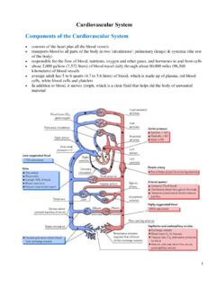
1 1 CardiovascularPathophysiology: left To Right ShuntsIsmee A. Williams, MD, Objectives Learn the relationships betweenpressure, blood flow, and resistance Review the transition from fetal tomature circulation Correlate clinical signs and symptomswith cardiac physiology as it relates toleft to Right shunt lesions: VSD, PDA, ASD Discuss Eisenmenger s Syndrome2 Pressure, Flow, Resistance Perfusion Pressure: Pressuregradient across vascular bed ! Mean Arterial - Venous pressure Flow: Volume of blood that travelsacross vascular bed Resistance: Opposition to flow Vessel diameter Vessel structure and organization Physical characteristics of bloodPoiseuille equationQ = !P"r4 R =8nl 8nl"r4!P = pressure dropr = radiusR = !Pn = viscosity Ql = length of tubeQ = flow3 Hemodynamics! PressureFlow (Q) =Resistance! PressureResistance = FlowTwo parallel fetal circulations!Placenta suppliesoxygenated blood viaductus venosus!Pulmonary blood flowminimal (<10%)!Foramen ovale directsductus venous blood toleft atrium (40%)!
2 Ductus arteriosusallows flow from PA todescending aorta (40%)4 Ductus Venosus and Streaming Ductus venosus diverts O2 bloodthrough liver to IVC and RA Amount varies from 20-90% Streaming of blood in IVC O2 blood from the DV!FO!LA!LV De-O2 blood from R hep, IVC !TV! RV SVC blood flows across TV!RV <5% SVC flow crosses FOO2 blood to high priority organs RV pumps De-O2 blood to PA!DA!DescAo ! lower body and placenta LV pumps O2 blood to AscAo!coronary + cerebral circ Aortic isthmus connects the twoseparate vascular beds5 Fetal Shunts Equalize Pressure RAp = LAp due to FO RVp = LVp due to DAUnlike postnatal life unless a largecommunication is work horse of fetal heart RV pumps 66% CO 59% goes to DA (88% RV CO) 7% goes to lungs (12% RV CO) LV pumps 34% CO 31% goes to AscAo Only 10% total COcrosses Ao isthmus6 Transition from Fetal to NeonatalCirculation Lose placenta "SVR Lungs expand mechanically "O2 vasodilates pulm vasc bed #PVR " PBF + "LA venous return "LAp DV constricts #RApThree Fetal Shunts Close LAp > RAp FO closure "O2 and # PGE1 DA and DV constrict RV CO # RV wall thickness # LV CO " LV hypertrophiesRV CO = LV COPostnatal circulation in series7 Regulation of Pulmonary Vascular Tone Vascoconstriction Hypoxia/acidosis High blood flow and pressure Failure of vessel maturation (noregression of medial hypertrophy)
3 Vasodilation Improved oxygenation Prostaglandin inhibition Thinning of vessel media(regression of medial hypertrophy)Fetal Pulmonary Vascular Bed Placenta is the organ of gas exchange Goal to bypass the fetal lungs Pulmonary Pressure >> Ao Pressure Low O2 tension causes Vasoconstriction Medial wall hypertrophy Pulmonary blood flow << Ao flow Pulmonary resistance >> Ao resistance Encourages shunting via DA to aorta8 Neonatal Pulmonary Vascular Bed Pulmonary Pressure # Ao Pressure Arterial vasodilation Medial wall hypertrophy persists Pulmonary Blood flow = Aortic Flow Ductus arteriosus closes Neonatal RV CO = LV CO Pulmonary resistance # Ao ResistanceAdult Pulmonary Vascular Bed Pulmonary Pressure << Ao Pressure 15 mmHg vs. 60 mmHg Arterial Vasodilation Medial wall hypertrophy regresses -remodeling Pulmonary Blood Flow = Aortic Flow Pulmonary Resistance << Ao Resistance Resistance = ! Pressure Flow9 Pulmonary Vascular Bed:Transition from Fetal to Adult!
4 PR=QRe-Cap: Fetal to Postnatal Fetus Shunts exist Lungs collapsed RV CO > LV CO (Parallel circ) Pulmonary pressure and resistance high Newborn Shunts close Lungs open RV CO = LV CO (Series circ) Pulmonary pressure and resistance drop10 left to Right Shunts Anatomic Communicationbetween Pulmonary andSystemic circulations Excess blood flow occurs fromthe Systemic ( left ) to thePulmonary ( Right ) circulationQp:Qs Extra flow is represented by theratio of pulmonary blood flow (Qp)to systemic blood flow (Qs) Qp:Qs = 1:1 if no Shunts Qp:Qs >1 if left to Right shunt Qp:Qs <1 if Right to left shunt Qp:Qs of 2:1 means pulmonaryblood flow is twice that of systemicblood flow11 Why do we care? Already oxygenated pulmonary venousblood is recirculated through the lungs Excess PBF causes heart failure (CHF) Size of the shunt and $ the amount ofPBF (Qp) determine how much CHF Shunt size determined by: Location of communication Size of communication Age of the patient Relative resistances to blood flow on eitherside of the communicationPulmonary Effects of L to R Shunt " PBF = " extravascular lung fluid transudation of fluid across capillariesfaster than lymphatics can clear Altered lung mechanics Tidal volume and lung compliance # Expiratory airway resistance " Pulmonary edema results if Qp andPulm Venous pressure very high Tachypnea12 Neurohumoral Effects of L to R Shunt Sympathetic nervous system andrenin-angiotensin system activation plasma [NE] and [Epi] " cardiac hormone B-type natriureticpeptide (BNP) " Tachycardia DiaphoresisMetabolic Effects of L to R Shunt Acute and chronic malnutrition Mechanism not clear " metabolic expenditures (" O2consumption) due to " respiratoryeffort and myocardial work # nutritional intake Poor growth/ Failure to thrive13 Pulmonary Hypertension.
5 End Stage " PBF causes sustained " PAp Pulm vascular bed fails to remodel Alveolar hypoxia may exacerbate Gradual effacement of the pulm arterioles Overgrowth of vascular smooth muscle Intimal proliferation Abnormal local vascular signaling Impaired endothelial function Pulm bed loses normal vasoreactivity fixed pulmonary HTN and irreversiblepulmonary vascular diseaseRe-Cap Flow, Resistance, Pressure Fetal and Transitional Circulation left to Right Shunts and CHF VSD PDA AVC ASD Eisenmenger14 Ventricular Septal Defect (VSD) left ventricle to Right ventricle Patent Ductus Arteriosus (PDA) Aorta to Pulmonary artery Atrioventricular Canal Defect (AVC) left ventricle to Right ventricle left atrium to Right atrium Atrial Septal Defect (ASD) left atrium to Right atrium Top 4 left to Right Shunt LesionsVSD most common CHD (20%) 2/1000 live births Can occur anywhere in the IVS Location of VSD has no effect on shunt Perimembraneous most common (75%) Muscular (15%) most likely to close Outlet (5%) most likely to involve valves " incidence in Asian pop (30%) Inlet (5%) assoc with AVC15 Ventricular Septal DefectVSD: Determinants of L to R shunt Size of VSD Difference inresistance betweenPulmonary andSystemiccirculations Difference inpressure betweenRV and LV16 VSD: Determinants of L to R shunt Small (restrictive) VSD: L to R shuntflow limited by size of hole Large (unrestrictive) VSD: L to R shuntflow is determined by Pressure andResistance If RVp < LVp, L to R shunt occurs If RVp = LVp, L to R shunt occurs ifpulmonary < aortic resistance Shunt flow occurs in systoleTransitional Circulation: Effects on L to R shunt in large VSD Fetus: bidirectionalshunt At Birth: No shunt Transition 1-7 wks PA/RVp $ to < LVp PA resistance $ to< Systemic L to R shunt %17 Large VSD: Hemodynamic Effects Flow LV!
6 RV! PA " Pulm Venous Return " LV SV initially byStarling mechanism " Pulm circ leads topulm vascular disease LA/LV volume overload " LV dilation leads tosystolic dysfxn & CHFVSD: Signs/Symptoms Asymptomatic at birth: PA = AoPressure and Resistance Signs of congestive heart failure aspulmonary pressure and resistance $ Poor feeding Failure to thrive (FTT) with preservedheight and low weight Tachypnea Diaphoresis Hepatomegaly Increased respiratory illness18 Harsh Holosystolic murmur loudest LLSB radiating to apex and back Smaller VSD = louder murmur Precordial Thrill 2 turbulence across VSD Mid-Diastolic rumble 2 " trans-Mitral flow LV heave 2 LV dilation Signs of CHF Gallop (S3), Hepatomegaly, Rales Signs of Pulm Vasc Disease #murmur, RV heave, loud S2, cyanosisVSD: Physical Exam CXR: Cardiomegally, "PVM Pulm Vasc Dz: large PAs EKG: LAE, LVH Pulm Vasc Dz: RVH ECHO: Location/Size VSD Amount/direction of shunt LA/LV size Estimation RV pressure CATH: only if suspect "PVR O2 step up in RVVSD: Laboratory Findings19 VSD: ElectrocardiogramVSD: CXR20 VSD: EchocardiogramVSD: Angiogram21 VSD: Management Does the patient have symptoms?
7 Size of the defect, RV/LV pressure,Pulm/Ao resistance Will the VSD close or #in size? Is there potential for complications? Valve damage, Pulm HTN Will the surgery be difficult? Will thesurgery be successful?VSD: Management Medical Digoxin Lasix Increased caloric intake 50% VSD size $ and CHF resolves Surgical Persistent CHF % pulmonary vascular resistance Valve damage Within first two years of life Catheter22 VSD: Endocarditis Prophylaxis Not for isolated VSD Yes for 1st 6 mo following repair ofVSD with prosthetic material ordevice Yes for life if there is a residualdefect at or adjacent to the site of aprosthetic device For dental and respiratory tractprocedures ONLY no longer for GI or GU proceduresPatent Ductus Arteriosus (PDA) Communication between Aortaand Pulmonary Artery 1/2500-5000 live births Risk factors: prematurity,rubella, high altitude23 PDA: Determinants of L to R shunt Magnitude L to R shunt depends on Length and diameter of ductus Relative resistances of Ao and PA " L to R shunt as Pulm resistance # Volume overload of PA, LA, LV Shunt flow occurs in systole and diastole PDA: Signs/Symptoms Small PDA: asymptomatic Large PDA.
8 CHF Diaphoresis Tachypnea Poor feeding FTT Hepatomegaly Respiratory infections Moderate PDA: Fatigue, Dyspnea,palpitations in adol/adults Afib 2 to LAE24 PDA: Physical Exam Continuous machine-like murmur atleft subclavian region Ao>PA pressure in systole anddiastole Congestive heart failurePDA: Laboratory Studies CXR: cardiomegally, " PVM EKG: LAE, LVH ECHO: measures size PDA, shuntand gradient, estimate PAp CATH: O2 step up in PA25 PDA: Management Indications for Closure CHF/failure to thrive Pulmonary hypertension Closure Methods Indomethacin if preemie Surgical ligation Transcatheter closure Coil DevicePDA Coil Closure26 Atrioventricular Canal Defect/Endocardial Cushion Defect Atrial SeptalDefect (Primum) Inlet VSD CommonAtrioventricularValveAVC: Management Closure always indicated Timing of surgery (elective by 6 mos.) Congestive Heart Failure Large left to Right shunt Mitral insufficiency Pulmonary hypertension Surgical repair ASD, VSD closure Repair of AV-Valves27 Summary: VSD, PDA and AVC Asymptomatic in fetus and neonate Progressive % in L to R shunt from 3-8wks of life as pulmonary pressureand vascular resistance $ Indications for intervention Congestive heart failure: FTT Pulmonary vascular disease End stage: Eisenmenger s syndromeAtrial Septum Formation Septum Primum grows downward Ostium Primum obliterates Fenestration in septum primumforms ostium secundum Septum secundum grows downwardand fuses with endocardial cushions Leaves oval-shaped opening Foramenovale Superior edge of septum primumregresses Lower edge becomes flap of FO28 Atrial Septal Defect Persistent communication between RAand LA Common: 1/1500 live births 7% of CHD Can occur anywhere in septum Physiologic consequences depend on.
9 Location Size Association with other anomaliesAtrial Septal Defect (ASD)29 ASD Types Ostium Secundum ASD (70%) 2:1 F>M Familial recurrence 7-10% Holt-Oram syndrome - upper limb defects Region of FO Defect in septum primum or secundum Ostium Primum ASD Inferior portion of septum Failure of fusion between septum primumand endocardial cushions Cleft in MV or CAVCASD Types Sinus Venosus ASD (10%) Incomplete absorption of sinus venosusinto RA IVC or SVC straddles atrial septum Anomalous pulmonary venous drainage Coronary Sinus ASD Unroofed coronary sinus Wall between LA and coronary sinusmissing Persistent L-SVC30 Patent Foramen Ovale Prevalence 30% of population Failure of fusion of septum primum andsecundum (flap of FO) Remains closed as long as LAp>RAp LAp<RAp Pulmonary HTN / RV failure Valsalva Paradoxical embolism and STROKEASD: Manifestations L to R shunt between LA and RA Amount of flow determined by: Size of defect Relative compliance of RV / LV Shunt flow occurs only in diastole L to R shunt " with age RV compliance " LV compliance # RA and RV volume overload31 ASD: Signs/Symptoms Infant/child usually asymptomatic DOE, fatigue, lower respiratory tractinfections Adults (prior age 40) Palpitations (Atrial tach 2 RAE) # stamina ( Right heart failure) Survival less than age-matched controls(5th-6th decade)ASD: Physical Exam Small for age Wide fixed split S2 RV heave Systolic murmur LUSB % flow across PV Mid-Diastolic murmur LLSB % flow across TV32 ASD: Laboratory Studies CXR: cardiomegally, " PVM EKG: RAD, RVH, RAE, IRBBB Primum ASD: LAD ECHO: RAE, RV dilation, ASD size,location, amount and direction ofshunt CATH: O2 step up in RAASD: Management Indications for closure RV volume overload Pulmonary hypertension Thrombo-embolism Closure method Surgical Catheter Delivered Device Cardioseal Amplatzer septal occluder33 Eisenmenger s Syndrome Dr.
10 Victor Eisenmenger, 1897 Severe pulmonary vascular obstruction 2 tochronic left to Right Shunts Pathophysiology High pulmonary blood flow& Shear Stress Medial hypertrophy + intimal proliferationleads to #cross-sectional area of pulm bed Perivascular necrosis and thrombosis Replacement of normal vascular architecture Pulmonary vascular resistance increases Right to left shunt Severe cyanosisMedial Hypertrophy34 Eisenmenger s SyndromeR to L flow via VSD Pressure: Pulmonary = Aortic Resistance Pulmonary > Aorta Blood flow: RV to LV Normal LA/LV size RV hypertrophy CyanosisEisenmenger s: Signs/Symptoms Infancy: CHF improves with # left to rightshunt Young adulthood: Cyanosis/Hypoxia: DOE, exerciseintolerance, fatigue, clubbing Erythrocytosis/hyperviscosity: H/A,stroke Hemoptysis 2 to infarction/rupturepulm vessels35 Eisenmenger s: Physical Exam Clubbing Jugular venous a-wave pulsations "RV pressure during atrial contraction Loud S2 RV heave (RV hypertension) Diastolic pulm insufficiency murmur No systolic murmurEisenmenger s: Lab findings No LV volume overload / " RV pressure CXR: Clear lung fields, prominent PAsegment with distal pruning, small heart EKG: RAE, RVH strain ECHO: RV hypertrophy, Right to left shuntat VSD, PDA, or ASD36 EKG: Eisenmenger s SyndromeEisenmenger s Syndrome: CXR37 Eisenmenger s: Management Avoid exacerbating Right to left shunt No exercise, high altitude, periph vasodilators Birth Control: 20-40% SAB, >45% mat mortality Medical Therapy: Pulmonary vasodilators.