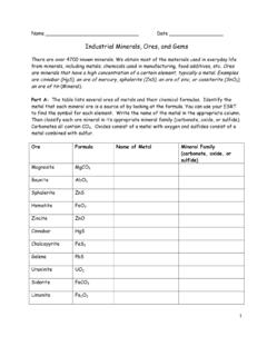
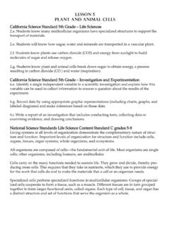
Transcription of Cardiovascular System Components of the Cardiovascular …
1 Cardiovascular System Components of the Cardiovascular System consists of the heart plus all the blood vessels transports blood to all parts of the body in two 'circulations': pulmonary (lungs) & systemic (the rest of the body). responsible for the flow of blood, nutrients, oxygen and other gases, and hormones to and from cells about 2,000 gallons (7,572 liters) of blood travel daily through about 60,000 miles (96,560. kilometers) of blood vessels average adult has 5 to 6 quarts ( to liters) of blood, which is made up of plasma, red blood cells , white blood cells and platelets In addition to blood, it moves lymph, which is a clear fluid that helps rid the body of unwanted material 1.
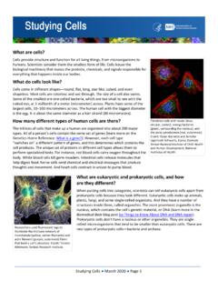
2 Anatomy of the Heart The heart is a muscular organ a little larger than your fist weighing between 7 and 15 ounces (200. to 425 grams). It pumps blood through the blood vessels by repeated, rhythmic contractions. The average heart beats 100,000 times per day pumping about 2,000 gallons (7,571 liters) of blood. The average human heart beating at 72 BPM (beats per minute), will beat approximately billion times during a lifetime of 66 years. The heart is usually situated in the middle of the thorax with the largest part of the heart slightly offset to the left underneath the breastbone or sternum and is surrounded by the lungs.
3 The sac enclosing the heart is known as the pericardium. The right side of the heart is the pulmonary circuit pump. Pumps blood through the lungs, where CO2 is unloaded and O2 is picked up. The left side of the heart is the systemic circuit pump. Pumps blood to the tissues, delivering O2 and nutrients and picking up CO2 and wastes. 2. Right Atrium: It collects deoxygenated blood returning from the body (through the vena cava). and then forces it into the right ventricle through the tricuspid valve. Left Atrium: It collects oxygenated blood returning from the lungs and then forces it into the left ventricle through the mitral valve.
4 The atrioventricular (AV) valves (Mitral & Tricuspid Valves) prevent flow from the ventricles back into the atria. Right Ventricle: It collects deoxygenated blood from the right atrium and then forces it into the lungs through the pulmonary valve. Left Ventricle: It is the largest and the strongest chamber in the heart. It pushes blood through the aortic valve and into the body. The pulmonary and aortic valves prevent back flow from the pulmonary trunk into the right ventricle and from the aorta into the left ventricle. Cardiac muscle cells are joined by gap junctions that permit action potentials to be conducted from cell to cell.
5 The myocardium also contains specialized muscle cells that constitute the conducting System of the heart, initiating the cardiac action potentials and speeding their spread through the heart. Aorta: It is the largest artery and carries oxygenated blood from the heart to the rest of the body. Superior Vena Cava: Deoxygenated blood from the upper parts of the body returns to the heart through the superior vena cava. Inferior Vena Cava: Deoxygenated blood from the lower parts of the body returns to the heart through the inferior vena cava. Pulmonary Veins: They carry oxygenated blood from the lungs back to the heart.
6 Pulmonary Arteries: They carry blood from the heart to the lungs to pick up oxygen. 3. External Heart Anterior View Frontal Section External Heart Posterior View 4. Pericardial Layers of the Heart Wall Epicardium visceral layer of the serous pericardium Myocardium cardiac muscle layer forming the bulk of the heart Fibrous skeleton of the heart crisscrossing, interlacing layer of connective tissue Endocardium endothelial layer of the inner myocardial surface Microscopic Anatomy of the Heart Muscle 5. Coronary Circulation Arterial Supply Venous Supply Pathway of Blood through the Heart and Lungs 6.
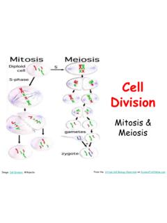
7 Heart Valves 7. Mitrial Valve Prolapse 8. Electrical System of the Heart 1. Sinoatrial Node (SA Node)-Pacemaker of the heart 2. Intra-atrial Pathway-carries electricity through atria 3. Internodal Pathway-carries electricity through atria 4. Atriaventricular Node (AV Node)-Back up pacemaker. Slows conduction 5. Bundle of His-last part of conduction in atria 6. Right Bundle Branch-carry electricity through R. Ventricle 7. Purkinje Fibers-distribute electrical energy to the myocardium 8. Left Bundle Branch-carries electricity through L. Ventricle Heartbeat Coordination Cardiac muscle cells must undergo action potentials for contraction to occur.
8 O The rapid depolarization of the action potential in atrial and ventricular cells (other than those in the conducting System ) is due mainly to a positive feedback increase in sodium permeability. o Following the initial rapid depolarization, the membrane remains depolarized (the plateau phase). almost the entire duration of the contraction because of prolonged entry of calcium into the cell through slow plasma-membrane channels. The SA node generates the current that leads to depolarization of all other cardiac muscle cells . o The SA node manifests a pacemaker potential, which brings its membrane potential to threshold and initiates an action potential.
9 O The impulse spreads from the SA node throughout both atria and to the AV node, where a small delay occurs. The impulse then passes in turn into the bundle of His, right and left bundle branches, Purkinje fibers, and nonconducting- System ventricular fibers. Calcium, mainly released from the sarcoplasmic reticulum (SR), functions as the excitation- contraction coupler in cardiac muscle, as in skeletal muscle, by combining with troponin. o The major signal for calcium release from the SR is calcium entering through voltage-gated calcium channels in the plasma membrane during the action potential.
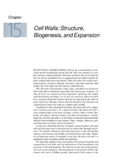
10 O The amount of calcium released does not usually saturate all troponin binding sites, and so the number of active cross bridges can be increased if cytosolic calcium is increased still further. Cardiac muscle cannot undergo summation of contractions because it has a very long refractory period. 9. Electrocardiogram (ECG or EKG) = record of spread of electrical activity through the heart P wave = caused by atrial depolarization (contraction). QRS complex = caused by ventricular depolarization (contraction) and atrial relaxation T wave = caused by ventricular repolarization (relaxation).