Transcription of Case 116 Sciatica with a sinister cause
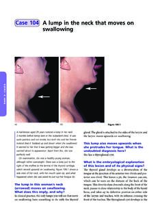
1 Part 2: cases241 Case 116 Sciatica with a sinister causeA retired railway worker aged 82 years was referred to the orthopaedic outpatient clinic with a note that read Severe lumbago and right-sided Sciatica , ? prolapsed intervertebral disc. Why did the surgeon discard the diagnosis of prolapsed intervertebral disc on reading the referral letter and even before seeing the old gentleman?A prolapsed intervertebral disc is prolapse of the nucleus pulposus, the jelly-like centre of the disc (Fig. ). This becomes less differentiated from the surrounding annulus fibrosus as age progresses. By the age of 55, it has more or less disappeared as a distinct structure.
2 A prolapsed disc in the late fifties is rare in the eighties it is an impossibility!When a detailed history was taken in the orthopaedic clinic, the patient described the pain as being in the region of the lumbar spine, the pelvis and spreading down the back of the thigh to the level of the ankle. It had commenced 3 or 4 months previously, and was getting much worse. It was a severe, dull pain, which kept him awake at night, was aggravated by coughing and straining, and was no longer relieved by analgesic tablets. In addition, the patient was having problems passing his urine, with hesitancy, poor stream, dribbling and a frequency of every 3 or 4 h during the day and three or four times at examination he was in obvious pain.
3 The lumbar spine was held rigid, with marked erector spinae spasm. Straight leg raising on the left was 70 , and on the right side 30 , and painful. There were no gross neurological anomalies. On rectal examination, the prostate was enlarged, irregular and woody is now your clinical diagnosis?The patient has marked prostatic symptoms and the clin-ical findings on rectal examination strongly suggest that he has a carcinoma of the prostate. The recent history of lumbar and pelvic pain, with sciatic radiation makes it likely that he has lumbar and pelvic secondary bone consultant urologist ordered X-rays of the chest, lumbar spine and pelvis.
4 The chest X-ray was clear. Figure is the film of the patient s lower lumbar spine, pelvis and hips. What does it demonstrate?There are extensive osteosclerotic deposits in the lumbar vertebrae, upper sacrum and the pelvic bones. Prostatic secondary deposits typically produce osteosclerotic lesions, whereas other secondary deposits are usually osteolytic (compare Case 100, p. 207).Give an anatomical explanation for the patient s lumbar pain and its radiationProstatic cancer spreads readily by Bateson s* valveless vertebral venous plexus from the prostatic venous plexus to the vertebrae and pelvic bones.
5 Wedging and patho-logical fractures account for the lumbar pain. Involve-ment of the sacral nerve roots explains the pain radiating down the back of the thigh (S2, S3, S4). Coughing and straining increase the spinal cerebrospinal fluid pressure and aggravate the may the diagnosis be confirmed in this patient?His prostate specific antigen was estimated and was 30 ng/ml, which is well in the range of disseminated prostatic cancer. Tissue diagnosis can be obtained by performing a transrectal biopsy of the prostate under ultrasound guidance. However, this is an invasive proce-dure, nearly always accompanied by marked haematuria *Oscar Bateson (1894 1979), Professor of anatomy, University of Pennsylvania, 2: cases242 Part 2: CasesFigure (a) Longitudinal section through the lumbar vertebrae showing a prolapsed intervertebral disc.
6 (b) MR image through a normal lumbar spine and sacrum. Note the excellent anatomical X-ray of the lower lumbar spine, pelvis and with the risk of septicaemia, such that it needs to be covered by broad spectrum antibiotics. In this case, it was considered that the diagnosis was sufficiently established to merit avoidance of a treatments are available for patients such as this with disseminated prostatic cancer? The mainstay of treatment is androgen suppression or the use of specific androgen antagonists, which will produce symptomatic relief in about 75% of cases. Radiotherapy may relieve the pain of bony deposits and can also be used for local control of the prostate to supplement hormonal therapy.
7 Urinary obstruction due to the prostatic tumour may resolve on hormonal therapy; if not, a transurethral endoscopic prostatectomy may be required.(a)(b)LigamentumflavumInterspin ous andsupraspinousligamentsAnnulus fibrosusNucleus pulposusA 'prolapsed disc'Intervertebral disc with central nucleus pulposusTermination of spinal cordSacral promontoryTermination of dural sacPosteriorlongitudinalligamentsAnterio r