Transcription of Case Report Mucinous myoepithelial carcinoma of the ...
1 Int J Clin Exp Pathol 2017;10(2) /ISSN:1936-2625/IJCEP0033885 case ReportMucinous myoepithelial carcinoma of the parotid gland: Report of a new histological variantShaodong Yang1,2, Ming Zeng1, Jiangli Zhang2, Xinming Chen21 The State Key Laboratory Breeding Base of Basic Science of Stomatology, (Hubei-MOST) & Key Laboratory of Oral Biomedicine Ministry of Education, School and Hospital of Stomatology, Wuhan University, Wuhan, Hubei, China; 2 Department of Pathology, School and Hospital of Stomatology, Wuhan University, Wuhan, Hubei, ChinaReceived June 17 2016; Accepted July 12, 2016; Epub February 1, 2017; Published February 15, 2017 Abstract: Mucinous myoepithelial carcinoma (MMC) is a newly recognized histologic variant of myoepithelial carci-noma, and only 14 cases have been reported in the English literature.
2 The authors herein Report an additional case of MMC of the parotid gland. A 72-year-old man presented with a right-sided facial mass. Computed tomography demonstrated a cm soft tissue mass in the right parotid, with peripheral enhancement and internal low-density. Total excision of the right parotid gland was performed. Histologically, the tumor was characterized by proliferation of predominantly mucin-containing signet-ring cells. Many polygonal to plasmacytoid cells with abun-dant eosinophilic, faintly granular cytoplasm were intermixed with the signet ring cells. Immunohistochemically, the tumor was diffuse positive for CK AE1/AE3, CK7, CK18, S100, and mammaglobin, but negative for CK14, CK20, vimentin, p63, p40, HHF-35, smooth muscle actin, calponin, DOG-1, Sox10, and Wilms tumor 1 gene protein.
3 The patient s postoperative course was uneventful and he was then lost to follow : myoepithelial carcinoma , myoepithelioma, Mucinous , signet-ring cell adenocarcinoma, signet-ring cell, parotid glandIntroductionMyoepithelial neoplasms are tumors composed almost exclusively of cells with myoepithelial differentiation. Most behave in a benign fash-ion and are designated myoepithelioma. Myo- epithelial carcinoma (MC), also known as malig-nant myoepithelioma, is the malignant counter-part of benign myoepitheliom, and is distin-guished from benign myoepithelioma by its infiltrative, destructive growth [1].
4 myoepithelial neoplasms account for about of all sali-vary tumors, and MC is even rarer, representing about 10% of myoepitheliomas [1-3]. However, myoepitheliomas show a wide spectrum of morphologic and immunophenotypic variation, resulting in difficulties in their diagnosis. They remain underrecognized and might not be as rare as has been reported. The tumor cells of myoepithelioms can be quite diverse, including spindled, stellate, epithelioid, plasmacytoid, basaloid, oncocytic, or clear cells [1-3]. In 2012, Esteva et al. reported 2 cases of unrecognized subtype of myoepithelial tumors that contained abundant intracellular mucin material, which they termed Mucinous myoepithelioma [4].
5 Bastaki et al. described the clinicopathologic, ultrastructural, immunophenotypic and molecu-lar features of four cases of primary signet ring cell adenocarcinoma, and designated them as secretory MC [5, 6]. According to the review by Gnepp, only 17 cases of Mucinous myopithelio-mas have been reported in the English litera-ture; Four have been classified as benign, and 13 as malignant neoplasms [7]. The authors herein Report an additional case of Mucinous myoepithelial carcinoma (MMC) of the parotid gland in a 72-year-old man, and discuss its clini-copathologic and immunohistochemical fea- reportA 72-year-old man presented with a right-sided facial mass.
6 He had first noticed a mass in the right parotid region 3 years earlier but left it Mucinous myoepithelial carcinoma of the parotid gland2225 Int J Clin Exp Pathol 2017;10(2):2224-2230untreated, and the mass had gradually enl- arged. The lesion had recently increased in size and associated with pain for the last 3 months. The patient reported a 30-year history of smok-ing (2 packs per day) and alcohol consumption (250 ml per day). The patient s past medical history included hypertension and hyperlipe-mia. His family history was unremarkable. Ph- ysical examination revealed a 6-cm, firm, and nodular mass in the right parotid region.
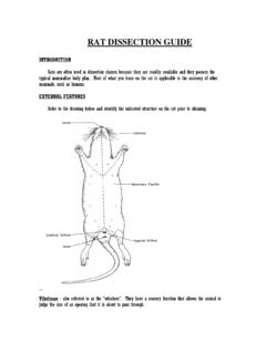
7 The swelling was vaguely palpable perorally, with a normal overlying mucosa. No skin abnormali-ties were noted. There was no facial palsy or palpable lymphadenopathy. Routine hemato-logic and biochemical examination, chest radio-graph, and abdomen ultrasound appeared nor-mal. Computed tomography (CT) with contrast demonstrated a cm, ill-defined soft tissue mass in the right parotid, with pe- ripheral enhancement and internal low-densi- ty (Figure 1). No abnormal lymphadenopathy was noted in the neck. Total excision of the right parotid gland was performed under a diag-nosis of a malignant parotid tumor, probably a carcinoma ex pleomorphic adenoma.
8 Frozen section histopathological evaluation showed a malignant tumor with features suggestive of acinic cell carcinoma . The patient s postopera-tive course was uneventful. He was then lost to follow examination of the surgical specimen demonstrated an ill-defined mass involving the entire parotid gland; the cut section was gray-ish white and gelatinous. Microscopic examina-tion revealed an unencapsulated neoplasm with infiltrative margins. Variably sized mucin lakes were divided into compartments by the fibrous bands and accounted for approximately two-thirds of the total tumor area, especially in the center (Figure 2A).
9 Tumor cells nests and single tumor cells were floating within the muci-nous material, resembling Mucinous (colloid) adenocarcinoma (Figure 2B). In other areas, the tumor was composed primarily of solid nests of pleomorphic cells separated by inter-vening fibrous stroma, which often had a multi-nodular architecture with hypercellular periph-eral rims and myxoid and necrotic central zones (Figure 2C). Most of the tumor cells were sig-net-ring cells, which demonstrated mucin-filled cytoplasmic vacuoles and displaced indented nuclei (Figure 2D). Both intracellular and extra-cellular mucins were strongly positive for muci-carmine and periodic acid Schiff after diastase staining.
10 In addition, many polygonal tumor cells with abundant eosinophilic, faintly granu-lar cytoplasm were intermixed with the signet ring cells (Figure 2E). Some of these tumor cells exhibited a plasmacytoid appearance, with ec- centric nuclei and abundant eosinophilic cyto-plasm. Tumor cells showed moderate to focally marked cellular pleomorphism with nuclear enlargement, vesicular or coarse chromatin, and variably prominent nucleoli (Figure 2F). Mitotic figures were frequently noted (24 mito-Figure 1. Axial (A) and coronal (B) computed tomography images show a large lesion with peripheral enhancement and internal low-density, located in the right parotid myoepithelial carcinoma of the parotid gland2226 Int J Clin Exp Pathol 2017;10(2):2224-2230 Figure 2.)