Transcription of Cat claw injuries in corneas of dogs and cats - Eye Vet
1 cat claw injuries in corneas of dogs and cats By: Natasha Mitchell MVB CertVOphthal MRCVS, Eye Vet, Crescent Veterinary Clinic, Dooradoyle Road, Limerick Cats have retractable claws which are versatile tools used for climbing, defence, balance, scratching and catching prey. They are also a formidable weapon which can inflict nasty scratches to animals perceived by the cat to be a threat (or a nuisance!). The most frequent victim is a younger dog, which is typically curious and has not yet learned to read hostile signals.
2 They also do not develop a menace response until 8-12 weeks of age, reducing the protection that would otherwise be afforded to their corneas by blinking at the appropriate moment and retracting the third eyelid, globe and head. Brachycephalic dogs have shallow orbits and more exposed eyes, therefore they are even more vulnerable to injury. Corneal lacerations from cat claw injuries are unfortunately relatively common. The prognosis for the condition depends on the severity of injury caused.
3 This in turn depends on the depth and angle of penetration, and the structures damaged. In general, there is a better prognosis for superficial (less than one third of the corneal thickness) rather than deep (greater than two thirds of the corneal thickness) corneal wounds, for peripheral rather than central corneal wounds, and for cats rather than dogs. Superficial ocular injuries carry a very good prognosis if the laceration is limited to the cornea, with no intraocular damage.
4 Deeper penetration of the claw may cause full thickness laceration of the cornea, with release of aqueous humour. Small perforations ( <1-2mm) may self-seal with a clot of aqueous humour and, later, fibrin. Larger perforations cause the sudden release of aqueous humour from the eye, which results in the iris moving forward to plug the deficit. This can result in anterior synechiae formation, or in prolapse of the iris, if the wound is sufficiently large.
5 If the defect is large, the iris may protrude through the corneal wound. Of key importance is whether the claw penetrates deep enough to rupture the anterior lens capsule. This leads to intraocular exposure to lens protein, which results in severe phacoclastic uveitis which is generally unresponsive to medical treatment. Very small lens capsule tears can result in cataract at the site which can later affect the entire lens. Other potential damage to the eye includes secondary reflex uveitis, hyphaema, corneal infection and intraocular infection (endophthalmitis).
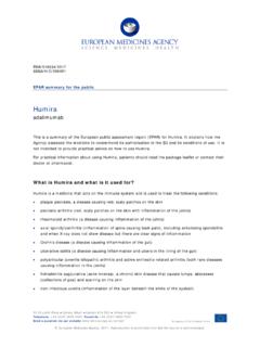
6 Infection is not common but needs to be prevented as the cat claw is potentially dirty. Evaluation: A careful assessment allows for the most appropriate treatment plan with the best possible outcome. The injured animal is usually uncomfortable and exhibits signs of ocular pain - blepharospasm, excessive lacrimation and third eyelid protrusion (Figure 1). In order to facilitate a more thorough examination and to provide pain relief for the patient, it is useful to apply topical anaesthetic, proxymetacaine , available as Minims single-use vials.
7 Topical anaesthetic should not be used as on-going treatment however, as it inhibits healing of the cornea. It is important to bear in mind that topical anaesthesia will reduce Schirmer tear test (STT) readings. The usual STT carried out in veterinary medicine measures both reflex and basal tears and is called the STT I. The STT II is measured after the application of topical anaesthetic and measures basal tear levels. Fig 1. A three-year-old Rottweiler on initial examination.
8 The eye was very painful and difficult to examine conscious. The eye was small due to hypotony, there was aqueous on the cornea and hyphaema. Gentle handling of the patient is necessary as even mild pressure could dislodge delicate temporary plugs sealing the injury, causing the wounds to open and intraocular contents to extrude. Sedation may be considered for un-cooperative patients to facilitate thorough examination. However it may result in the eye rolling downwards, which will hinder the examination.
9 Therefore if direct examination is not possible despite application of topical anaesthetic and careful restraint, general anaesthesia is better advised. The greater relaxation provided minimises the risk of further corneal damage or rupture, and the eye can be more easily manipulated into a forward-gazing position for examination by means of grasping the conjunctiva with forceps or placing conjunctival perilimbal stay sutures. Sedation and anaesthesia (depending on the choice of drug) typically reduce both the STT readings and IOPs.
10 Drugs which increase intraocular pressure (IOP) either directly or indirectly should be avoided for example ketamine and morphine. There may be scratch marks on the face, eyelids or third eyelid. Every case with such injuries should be carefully assessed for corneal involvement. Similarly, patients with corneal laceration should have the integrity of the adnexal structures examined to investigate the extent of the injuries . Examination behind the third eyelid and in the conjunctival fornices is indicated to ensure that part of the claw has not been left behind.