Transcription of Cell Proliferation Assay and Staining Protocol - …
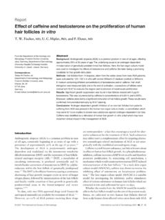
1 4 Measuring Cell Viability / CytotoxicityIntroductionCell viability and cytotoxicity assays are used for drug screening and cytotoxicity tests of chemicals. Fig. 1 indicates various reagents used for cell viability detection. They are based on various cell functions such as enzyme activity, cell membrane permeability, cell adherence, ATP produc-tion, co-enzyme production, and nucleotide uptake activity. Many have established methods such as Colony Formation method, Crystal Violet method, Tritium-Labeled Thymidine Uptake method, MTT, and WST methods, which are used for counting the number of live cells. Trypan Blue is a widely used Assay for Staining dead cells. In this method, cell viability must be determined by counting the unstained cells with a microscope or other instruments.
2 However, Trypan Blue Staining cannot be used to distinguish between the healthy cells and the cells that are alive but losing cell functions. In the Colony Formation method, the number of cell colonies are counted using a microscope as a cell viability indicator. In the Tritium-Labeled Thymidine Uptake method, [3H]-thymidine is involved in the cell nucleus due to the cell growth, and the amount of the tritium in the nucleus is then measured using a scintillation counter. Though the Tritium-labeled thymidine uptake Assay is sensitive to determine the infl uence on the DNA polymerization activity, it requires radioisotope which causes various concerns. The 51Cr method is highly sensitive, and is commonly used to determine low levels of cytotoxicity.
3 However, the use of 51Cr also causes problems in handling, storage, and disposal of the material. Cellular enzymes such as lactate dehydrogenase, adenyl-ate kinase, and glucose-6-phosphate dehydrogenase are also used as cell death markers, and there are several viable cellCalcein-AM(non-fl uorescent compound)esterasenucleusCalceinmitochond riaATPNADH, NADPH/DehydrogenaseWST-8 formazan dyeWST-8 (colorless substrate)MTTMTT formazan dyes (crystals)Luciferase, O2(bioluminescence)Fig. 1 Reagents for cell viability detection[3H]ThymidineDNA Polymeraseproducts available on the market. However, adenylate kinase and glucose-6-phosphate are not stable and only lactate dehy-drogenase does not lose its activity during cell death assays. Therefore, cell death assays based on lactate dehydrogenase (LDH) activity are more reliable than other enzyme-based cell death methods using MTT and WST rely on a reduc-tive coloring reagent and dehydrogenase in a viable cell to determine cell viability with a colorimetric method.
4 This method is far superior to the previously mentioned methods because it is easy-to-use, safe, has a high reproducibility, and is widely used in both cell viability and cytotoxicity tests. Therefore, this method is suitable for those who are just beginning such experiments. Among the enzyme-based assays, the MTT as-say is the best known method for determining mitochondrial dehydrogenase activities in the living cells. In the method, MTT is reduced to a purple formazan by NADH. However, MTT formazan is insoluble in water, and it forms purple needle-shaped crystals in the cells. Therefore prior to measuring the absorbance, an organic solvent is required to solubilize the crystals. Additionally, the cytotoxicity of MTT formazan makes it diffi cult to remove cell culture media from the plate wells due to fl oating cells with MTT formazan needles, giving signifi cant well-to-well error.
5 Dojindo developed highly water-soluble tetrazolium salts called WSTs. WSTs produce water-soluble formazans and are suitable for cell Proliferation and cytotoxicity assays. WSTs receives two electrons from viable cells to generate a yellow, orange, or purple formazan dye. WST-8, a highly stable WST, is utilized in Cell Counting Kit-8 (CCK-8). The electron mediator used in this kit, 1-Methoxy PMS, is also highly stable. Therefore, CCK-8 is stable for at least 6 months at the room temperature and for one year at 0-5 oC. Since WST-8, WST-8 formazan, and 1-Methoxy PMS have no cytotoxicity in the cell culture media, additional experiments may be carried out using the same cells from the previous assays refl ect cell conditions with more sensitivity than the other assays because they depend on several elements including dehydrogenase, NAD(H), NADP(H), and mitochondrial activity.
6 The major diff erence between CCK-8 and the MTT Assay , other than MTT s toxicity, is the enzymes involved. The CCK-8 Assay involves most of the dehydrogenase in a cell. On the other hand, MTT only involves mitochondrial dehydrogenase. Therefore, the MTT Assay depends on mitochondrial activity, not the cell itself. Additionally, CCK-8 is far more sensitive than the MTT Assay . Since WST-8 formazan is water soluble, it does not form crystals like MTT. Therefore, after 1-4 hours of incubation with the CCK-8 solution, measurement of at 450 nm gives the number of viable cells. No extra steps are Request Available (US/Canada Only) Cell Counting Kit-8 - Ready-to-use one solution - Stable for 12 months when stored at 4 oCDevices, Tools Microplate Reader with a 450 - 490 nm fi lter 96 well microplate, sterilized clear plate for cell Assay Multi-channel pipette (8 or 12 channel: 10-100 l) Pipette tips for 10-100 l CO2 incubator Clean bench Hematocytometer or cell counter Centrifuge and rotor for a 15 ml centrifuge tubeReagents Cell Counting Kit -8 [product code: CK04] Cell culture media Material to be tested PBS or other buff ers for the preparation of material solutions if cell culture medium cannot be MaterialsProduct InformationMeasuring Cell Viability/Cytotoxicity.
7 Cell Counting Kit-8 Product DescriptionCell Counting Kit-8 is a colorimetric Assay for the determination of viable cell numbers and can be used for cell Proliferation assays as well as cytotoxicity assays. Cell Counting Kit-8 uses a tetrazolium salt, WST-8, which produces the water soluble WST-8 formazan. Since this orange colored formazan does not require dissolving, no solubilizing process is required. Re-sults are obtained after 3 simple steps: 1) add, 2) incubate, and 3) read. This kit is applicable for 96-well microplate assays and can also be applied to High-Throughput Screening such as a 384-well microplate. WST-8 is not cell permeable, which results in low cytotoxicity. Therefore after the Assay it is possible to continue further experiments using the same : cell counting, cell Proliferation experiments, cytotoxicity tests, drug sensitivity testsIf you use Cell Counting Kit-8 frequently, store in a refrigerator.
8 The solution is stable for one year at 4 oC. The solution is also stable at room temperature for 6 you plan not to use the Cell Counting Kit-8 for more than a year, aliquot the Cell Counting Kit-8 solution and store in a freezer at -20 oC to avoid repeated freeze and Counting Kit-8 Product codeUnitComponentsCK04-01 100 tests 1 ml bottle x 1CK04-05 500 tests 5 ml bottle x 1 CK04-111,000 tests5 ml bottle x 2CK04-133,000 tests5 ml bottle x 6CK04-2010,000 tests 100 ml bottle x 1 One test corresponds to one well of the 96-well the cells to be assayed from a culture fl ask. Adjust the con-centration of the cell suspension to 5x10⁴ cells per ml using a hemato-cytometer or cell Serial Dilution a. Using an 8 channel multi-pipette, add 100 l of media to each well of a 96 well microplate.
9 B. Add 100 l of a 5x10⁴ cells per ml solution to the wells of fi rst trip- licate row. c. Take 100 l from the fi rst triplicate row, add it to the next well and mix. The process is repeated as indicated in the fi gure 2. Reserve the fi nal well for the negative control (Blank). This well should contain media only (no cells) for measurement of the the plate in a CO2 incubator. Cells in the log (dividing) phase is strongly recommended. <For adherent cells> Pre-incubation is required for 2-4 hours in order to attached the cells to the plate. <For suspension cells or adherent cells that do not need to be adhered> Pre-incubation can be skipped. Most cell lines would be in the log phase after 48-72 hours of 10 l of Cell Counting Kit-8 reagent to each well on the 96-well in a CO2 incubator for 1-4 hours to an absorbance on a microplate reader using a 450 nm fi lter.
10 Establish your standard curve by plotting the number of cells on the x -axis and the absorbance on the adherent cells, recover the cells using trypsin to detach cells, and use a cell scraper if to experimental example on the next page for instruction on serial using a plate or petri dish other than a 96 well plate, please add reagent equal to 1/10 the media volume. Due to the low volume of reagent added, it is recommended to place the pipette tips against the well wall to add the reagent. If the reagent sticks to the well wall, tap the plate lightly to mix with the performing the experiment for the fi rst time, we recommend taking readings every hour at 1, 2, 3 and 4 the amount of formazan produced will differ with each cell types, the amount of coloration will diff er even if the time between adding the reagent and taking a reading is the same.