Transcription of CENTERPIECE PLATE FIXATION SYSTEM - MT Ortho
1 CENTERPIECE PLATE FIXATION SYSTEMS urgical TechniqueCENTERPIECE Surgical Technique1 Dear Colleagues:The preferred surgical procedure for the treatment of multi-level cervical myelopathy remains a matter of discus-sion. In selecting a procedure to recommend to their patients, surgeons must account for a number variables, such as the sources and location of compression, cervical alignment, segmental instability patterns, comorbid medical conditions, functional requirements of the host, degree of preoperative axial pain, etc. Laminoplasty has been proven to yield the same neurologic outcomes as multi-level anterior decompression and fusion procedures. However, the complication rates are lower with laminoplasty, and motion is preserved rather than sacrificed.
2 Early active range of motion may also be encouraged, provided the position of the laminae is secured during myriad of laminoplasty techniques have been described, none of which appear to differ to any great extent in their success rates. The choice of which method a surgeon might use appears to be primarily a matter of training and familiarity, as are the details of how the laminae are maintained in their open position. The principle school of thought described in this technique is the Open Door procedure. We have worked closely with Medtronic Sofamor Danek to develop a SYSTEM of implants to facilitate laminar FIXATION during Open Door laminoplasty monograph concerns the use of the CENTERPIECE PLATE FIXATION SYSTEM with and without an interpositional allograft.
3 We hope that you will note its potential advantages for your patients: Anatomic contouring Ease of application Secure laminar FIXATION Adaptability to procedures with and without allograft Facilitation of early active motion and rehabilitationFor those familiar with laminoplasty, we think that you will find the CENTERPIECE PLATE FIXATION SYSTEM a welcome enhancement to your technique. For those considering adding laminoplasty to their surgical repertoire, we think that the CENTERPIECE SYSTEM will enhance the appeal of the procedure and make it easier to ,John G. Heller, MD Jeffrey C. Wang, MDPrefaceCENTERPIECE Surgical Technique2 Implant Features ..3 Instruments ..5 Overview.
4 6 Patient Postioning ..7 Surgical Exposure ..8 Open Door Technique Open Side Trough Preparation ..9 Hinge Side Trough Preparation .. 10 Opening the Laminoplasty .. 11 Keeping the Door Open A. Utilizing the Open Door PLATE .. 12 B. Utilizing the Graft PLATE .. 15 Use of Lateral Hole PLATE .. 17 Use of Wide Mouth PLATE .. 18 Use of Hinge PLATE .. 19 Implant Removal .. 20 Product Ordering Information .. 21 Important Information on the CENTERPIECE PLATE FIXATION SYSTEM .. 22 Table of ContentsCENTERPIECE Surgical Technique3 Open Door Plates Pre-cut, pre-contoured PLATE design Laminar shelf of PLATE allows for easy FIXATION to lamina Multiple screw hole options for flexibility in screw placement Intrinsic stability provided by design of PLATE Kickstand design of PLATE aids in stability when placed on lateral mass Color coded Available in 8mm 18mm sizes in 2mm increments Pre-cut.
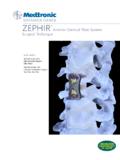
5 Pre-contoured PLATE design Oval shaped center screw hole in the graft PLATE allows for fine adjustments of the PLATE on the allograft Multiple screw hole options for flexibility in screw placement Color coded Available in 8mm 18mm sizes in 2mm increments Medial/lateral orientation of the lateral mass screw holes allows for flexible screw placement in the event that the surface area of the lateral mass has been reduced in its cranial-caudal dimension, especially following supplemental foraminotomies Color coded Available in 8mm 18mm sizes in 2mm incrementsGraft PlatesLateral Hole Plates (in OD and GP)Implant FeaturesCENTERPIECE Surgical Technique4 Small angled PLATE designed to secure a floppy or displaced hinge Wider laminar shelf used to accomodate thick laminae Color coded Available in 8mm - 18mm sizes in 2mm increments Self-tapping Precision instrumentation with stab-and-grab feature Color coded Available in and diameters, 5mm - 11mm lengths in 2mm increments All cortical graft for added stability Simplified surgical technique by eliminating the need to shape resected autograft Curved edges of allograft provide a secure fit between the lamina and lateral mass Pre-drilled center screw hole allows for immediate access for screw insertion.
6 Eliminating a procedural step Freeze-dried Available in 8mm 18mm sizes in 2mm incrementsHinge PlatesWide Mouth Plates (OD Lateral Hole PLATE and OD Standard Hole PLATE )Bone ScrewsAllograftTop ViewSide ViewImplant FeaturesCENTERPIECE Surgical Technique5 Universal handleBone TrialPlate HolderScrewdriver SleeveBone CutterDrill Bit with x StopScrewdriver ShaftInstrumentsCENTERPIECE Surgical Technique6 Cervical laminoplasty can achieve spinal cord decompression in cases of multi-level myelopathy or myeloradiculopa-thy due to cervical spondylosis, ossification of the posterior longitudinal ligament (OPLL), and similar conditions. The most common reason for failure of laminoplasty has been re-stenosis due to hinge closure.
7 Various techniques have been employed to hold the door open while the host heals the laminar hinge in the expanded position. Ideally, a method of achieving laminar FIXATION should be technically intuitive, provide secure laminar FIXATION , while minimizing the risk of iatrogenic injuries, blood loss and operative time. The authors describe the use of the novel CENTERPIECE PLATE FIXATION SYSTEM designed to accomplish these goals during Open Door laminoplasty. The technical issues relevant to performing the laminoplasty and securing the laminae are discussed. The plates have been proven to be biomechanically equivalent to the currently-used techniques. The use of these plates will potentially allow the patient to engage in an early active rehabilitation protocol while minimizing the risk of re-stenosis of the canal.
8 This may ultimately lead to better preservation of motion and decreased axial neck pain following Surgical Technique7 The patient is positioned prone as for most other posterior cervical procedures, with the head secured in a Mayfield three-pin head-holder, preferably in slight flexion (Figures 1 and 1a). Some cervical flexion helps reduce the overlap of the laminae and facet joints, which facilitates the laminoplasty itself. A reverse Trendelenberg position may help decrease bleeding from epidural and paravertebral 1 Figure 1aPatient PositioningCENTERPIECE Surgical Technique8 The surgeon performs a midline posterior exposure from the inferior aspect of C2 to the superior aspect of T1 (Figures 2 and 3). The lateral dissection follows the subperiosteal plane out to the mid-portion of the lateral masses.
9 Unlike the exposure required for a laminectomy and fusion, the muscle origins and insertions over the lateral half of the lateral masses are preserved. The insertion of the extensor muscles is only detached from the lower laminar margin of C2 to afford access to the C2-3 inter-laminar space. The junction of the medial aspect of the lateral mass with the lateral portion of the lamina is identified at each level planned in the decompression. At this point it is particularly helpful to correlate the local surface anatomy with the preoperative axial images. Figure 2 Figure 3 Note: In the event that the posterior decompression ought to extend to the C2 level, this can be accom-plished while respecting the integrity of the C2 posterior arch and the majority of its muscular origins and insertions.
10 A so-called dome laminectomy is performed by using a burr and kerrison to remove the lower margin of C2, followed by the cancellous bone and ventral cortex (Figures 4, 4a and 4b).Figure 4 Figure 4aFigure 4bSurgical ExposureCENTERPIECE Surgical Technique9 Open Side Trough PreparationThe open side trough is prepared with a burr along the junction of the lamina and the lateral mass. Three layers of bone must be removed in turn: the dorsal cortex, followed by the cancellous layer and then the ventral cortex (Figure 5 and 5a). Hemostasis of the bone surfaces can be achieved with the use of thin bone wax match sticks or applying a slurry of powdered GelFoam and thrombin solution. The completion of the bone separation on the open side can be performed with a millimeter Kerrison Rongeur.