Transcription of CHAPTER 1
1 CHAPTER 1 dental RADIOLOGYINTRODUCTIONThe purpose of dental radiography is to recordimages of a patient's oral structures on film by usingX-rays. When the X-ray films are processed, theresulting radiographs provide the dental officer with avaluable diagnostic aid. In the case of death,radiographs can be used to aid in identification asdiscussed in CHAPTER 10, "Forensic Dentistry."The first section of this CHAPTER covers the basicfundamentals of dental radiography . Included are thephysics and biology of X-radiation can be harmful, you mustobserve certain safety precautions when using anX-ray machine or working in an area where one isbeing used.
2 These precautions are covered in thesecond part of this major portion of this CHAPTER is devoted toexplaining how to operate a dental X-ray machine,expose intraoral radiographs (radiographs taken insidethe patient s mouth), process the X-ray films, andmount the finished last part of this CHAPTER covers the panoramicX-ray machine, which you will use to make extraoralradiographs (radiographs made outside the patient smouth).FUNDAMENTALS OF DENTALRADIOLOGYOral radiography is the art of recording images of apatient s oral structures on film by using X-rays(roentgen rays). The rays were recognition ofWilhelm Konrad Roentgen, a scientist, who firstdiscovered X-rays in 1895.
3 While experimenting witha device called a Crookes tube, which generatedcathode rays, he noted that a photographic platecompletely wrapped in black paper and lying near thetube was fogged when developed. He realized thatsome form of invisible ray, able to pass through theblack paper, must be coming from the tube. Later,while in his darkened laboratory, he noticed that afluorescent screen located six feet away was knew that the cathode rays could travel only shortdistances outside the cathode tube and realized he was1-1observing a new, unknown ray, which he called anX-ray because the symbol "X" is used for theunknown in first dental radiograph was taken the sameyear by Dr.
4 Otto Walkoff. Within 10 years,radiographs were being used for diagnosis of medicaland dental conditions, for X-ray therapy, and- forscientific studies. Although technology over the yearshas made tremendous improvements in X-rayequipment, the basic concepts are the visible light rays, X-rays are electromagneticrays that travel in a wave motion. The measurement ofthis wave motion is called a wavelength. The basicdifference between X-rays and other electromagneticrays is in their wavelength. X-rays have an extremelyshort wavelength, which enables them to penetratematter that usually absorbs or reflects light or otherelectromagnetic rays with longer X-rays share the properties of otherelectromagnetic rays, their action is considerablydifferent.
5 Some of the characteristics and properties ofX-rays are:They travel in straight lines at the speed of affect photographic film by producing ahidden image made visible by cause certain substances to fluoresce(glow).They cause irritation of living cells and, in largeamounts, can cause necrosis (death) of the cells,a fact that necessitates caution in using are produced when a metal (tungsten)target is bombarded by a stream of electrons. TheX-rays are emitted in the tubehead and directed by thetubehead cone through the subject, producing animage on the density of the X-ray image is controlled byfour factors: kilovoltage (kVp), exposure time,milliamperage (mA), and target-film distance (TFD).
6 All of these factors are interrelated and may be variedby the operator. The procedures for setting thesefactors will be discussed SAFETYP roper safety precautions must be observed by allpersons working in or near an area where X-rays arebeing generated. X-rays can be dangerous. Long termoverexposure to radiation may result in loss of hair,redness and inflammation of the skin, blood countchange, cell atrophy (wasting away), ulcerations,sterility, genetic damage, cancer, leukemia, and are safety measures designed to protect thepatient and the health care team from the dangers ofoverexposure to radiation and the operation of X-rayequipment.
7 You must observe these safety measureswhen working in radiology . Your command will haveinstructions and standard operating procedures (SOP)for the operation of dental radiographic (X-ray) unitsand equipment. You will be required to read theseprocedures if you are newly assigned to the radiologydepartment. There are other numerous responsibilitiesthat include providing radiology support for oraldiagnosis, log maintenance, infection control, testingfor quality control, and processor PROTECTIONA number of precautions are taken to prevent thepatient from being exposed to inappropriate diagnosticradiation. The decision to order dental radiographs isdetermined by the dental officer on a case by case basisfor each patient.
8 Only a dental officer is authorized toorder and diagnostically interpret dental the most important safety measure is theresponsibility of the assistant: When takingradiographs, you should always have patients wearlead aprons and thyroid collars to shield theirreproductive organs and thyroid glands. There isonly one exception to this rule; when obtaining apanorex radiograph, the thyroid collar is not usedsince it blocks part of the X-ray beam. In addition,always ask a female patient whether or not she ispregnant or if pregnancy is questionable, before takingradiographs. If she is pregnant, consult the radiation safety measures include X-raymachines that have built-in safeguards that filter outharmful radiation and restrict the central X-ray to thesmallest possible area.
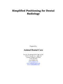
9 Fast film is used to shortenexpos ure time; and only essential radiographs aretaken on 1-1. Environmental dosimetry radiation film PROTECTIONWhen you work near a source of radiation, yourX-ray department will be issued an environmentaldosimetry radiation film badge (fig. 1-1).feet from the tube head and never in the direct line ofradiation during expos ure. These film badgesAppropriately placed environmental filmbadges are used to monitor stray radiation that mayoccur in and around the X-ray department. Thebadges are placed in the X-ray room behind thetechnicians protective lead-lined barrier or at least 6contain X-ray sensitive film in a light-tight film packets are collected every 6 to 7 collection, the film is sent to the radiationdetection laboratory for processing and abnormally high readings ( , greater REM [Radiological Equivalent Mammel])shall be referred to the Radiation Health Office you take radiographs on a patient, observethe following precautions to avoid unnecessaryexposure to radiation.
10 NEVER stand in the path of the central X-raybeam during hold the X-ray film packet in thepatient's mouth during hold the tube head or the tube headcylinder of the X-ray machine during stand behind a lead-lined screenduring an FILM LOGA nother portion of radiation safety is to accountfor all radiographs that are taken. An X-ray film log1-2shall be maintained in all X-ray rooms and will containthe following information:Column 1:Patient's NameColumn 2:Patient's SSNC olumn 3:Patient's UnitColumn 4:Rank/Rate/Retired/ 5:Number of X-ray exposures andtype: bitewing, periapical, occlusal,panographColumn 6:kVp, mA, exposure timeColumn 7:Reason retake X-ray required (ifapplicable)When stating the reason for a retake X-ray, bespecific on the nature of the retake, for example:conecut, elongated, foreshortened, dark image, X-RAY MACHINESThe most commonly used X-ray machine is thewall-mounted dental X-ray unit (fig.)