Transcription of Chapter 13 - Normal Labor and Delivery
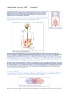
1 267 Chapter 13 Normal Labor and DeliverySarah Kilpatrick and Etoi GarrisonLABOR: DEFINITION AND PHYSIOLOGYL abor is defined as the process by which the fetus is expelled from the uterus. More specifically, Labor requires regular, effective contractions that lead to dilation and effacement of the cervix. This Chapter describes the physiology and Normal characteristics of term Labor and physiology of Labor initiation has not been com-pletely elucidated, but the putative mechanisms have been well reviewed by Liao and Labor initia-tion is species-specific, and the mechanisms in human Labor are unique. The four phases of Labor from quiescence to involution are outlined in Figure The first phase is quiescence and represents that time in utero before Labor begins when uterine activity is suppressed by the action of progesterone, prostacyclin, relaxin, nitric oxide , parathyroid hormone related peptide, and possibly other hormones.
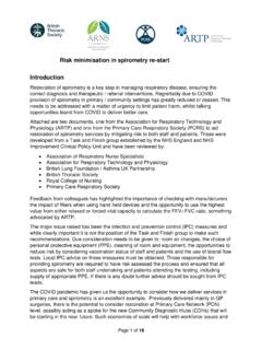
2 During the activation phase, estrogen begins to facilitate expression of myometrial receptors for prosta-glandins (PGs) and oxytocin, which results in ion channel activation and increased gap junctions. This increase in the gap junctions between myometrial cells facilitates effec-tive In essence, the activation phase readies the uterus for the subsequent stimulation phase, when uterotonics, particularly PGs and oxytocin, stimulate regular contractions. In the human, this process at term may be protracted, occurring over days to weeks. The final phase, uterine involution, occurs after Delivery and is mediated primarily by oxytocin. The first three phases of Labor require endocrine, paracrine, and autocrine interac-tion between the fetus, membranes, placenta, and fetus has a central role in the initiation of term Labor in nonhuman mammals; in humans, the fetal role is not completely understood (Figure 13-2).
3 2-5 In sheep, term Labor is initiated through activation of the fetal hypothalamic-pituitary-adrenal axis, with a resultant increase in fetal adrenocorticotrophic hormone and ,5 Fetal cortisol increases production of estradiol and decreases production of progesterone by a shift in placental metabolism of cor-tisol dependent on placental 17 -hydroxylase. The change in the progesterone/estradiol ratio stimulates placental pro-duction of oxytocin and PG, particularly PGF2 .4 If this increase in fetal adrenocorticotrophic hormone and cortisol is blocked, parturition is In contrast, humans lack placental 17 -hydroxylase and there is no increase in fetal cortisol near term. Rather, in humans, uterine activation may be potentiated in part by increased fetal adrenal pro-duction of dehydroepiandrostenedione, which is converted in the placenta to estradiol and estriol.
4 Placental estriol stimulates an increase in maternal (likely decidual) PGF2 , PG receptors, oxytocin receptors, and gap junctions. In humans, there is no documented decrease in progesterone near term and a fall in progesterone is not necessary for Labor initiation. However, some research suggests the possibility of a functional progesterone withdrawal in humans: Labor is accompanied by a decrease in the con-centration of progesterone receptors, as well as a change in the ratio of progesterone receptor isoforms A and B in both the myometrium6,7 and More research is needed to elucidate the precise mechanism through which the human parturition cascade is activated. Fetal maturation may play an important role, as well as maternal Labor .
5 Definition and Physiology 267 Mechanics of Labor 268 Uterine Activity (Powers) 270 The Fetus (Passenger) 270 The Maternal Pelvis (Passage) 272 Cardinal Movements in Labor 275 Engagement 275 Descent 277 Flexion 277 Internal Rotation 277 Extension 277 External Rotation 277 Expulsion 277 Normal Progress of Labor 277 Interventions Affecting Normal Labor Outcomes 279 Active Management of Labor 279 Second Stage of Labor 280 Spontaneous Vaginal Delivery 280 Delivery of the Placenta and Fetal Membranes 281 Episiotomy, Perineal Injury, and Perineal Repair 281 KEY ABBREVIATIONSA merican College of Obstetricians and Gynecologists ACOGC ephalopelvic Disproportion CPDLeft Occiput Anterior LOAO cciput Anterior OAOcciput Posterior OPOcciput Transverse OTProstaglandins PGsRandomized Controlled Trial RCTR ight Occiput Anterior ROA268 Section III Intrapartum Carehigh-affinity oxytocin receptors have also been isolated from human amnion and decidua parietalis but not decidua ,13 It has been suggested that oxytocin plays a dual role in parturition.
6 First, through its receptor, oxytocin directly stimulates uterine contractions. Second, oxytocin may act indirectly by stimulating the amnion and decidua to produce ,16,17 Indeed, even when uterine contractions are adequate, induction of Labor at term is successful only when oxytocin infusion is associated with an increase in PGF binding to its receptor activates phospolipase C. In turn, phospholipase C increases intracellular calcium both by stimulating the release of intracellular calcium and by promoting the influx of extracellular calcium. Oxytocin stimulation of phospholipase C can be blocked by increased levels of cyclic adenosine monophosphate. Increased calcium levels stimulate the calmodulin-mediated activa-tion of myosin light-chain kinase.
7 Oxytocin may also stimulate uterine contractions via a calcium-independent pathway by inhibiting myosin phosphatase, which in turn increases myosin phosphorylation. These pathways (PGF2 and intracellular calcium) have been the target of multiple tocolytic agents: indomethacin, calcium channel blockers, beta mimetics (through stimulation of cyclic adenosine monophosphate), and OF LABORL abor and Delivery are not passive processes in which uterine contractions push a rigid object through a fixed aperture. The ability of the fetus to successfully negotiate the pelvis during Labor and Delivery depends on the complex interactions of three variables: uterine activity, cues that affect circadian cycling. There are distinct diurnal patterns of contractions and Delivery in most species, and in humans, the majority of contractions occur at ,9 Oxytocin is used commonly for Labor induction and aug-mentation; a full understanding of the mechanism of oxy-tocin action is helpful.
8 Oxytocin is a peptide hormone synthesized in the hypothalamus and released from the posterior pituitary in a pulsatile fashion. At term, oxytocin is a potent uterotonic agent that is capable of stimulating uterine contractions at intravenous infusion rates of 1 to 2 Oxytocin is inactivated largely in the liver and kidney, and during pregnancy, it is degraded primarily by placental oxytocinase. Its biologic half-life is approxi-mately 3 to 4 minutes, but appears to be shorter when higher doses are infused. Concentrations of oxytocin in the maternal circulation do not change significantly during pregnancy or before the onset of Labor , but they do rise late in the second stage of ,11 Studies of fetal pitu-itary oxytocin production and the umbilical arteriovenous differences in plasma oxytocin strongly suggest that the fetus secretes oxytocin that reaches the maternal side of the ,12 The calculated rate of active oxytocin secretion from the fetus increases from a baseline of 1 mIU/min before Labor to around 3 mIU/min after spon-taneous differences in myometrial oxytocin receptor distribution have been reported.
9 With large numbers of fundal receptors and fewer receptors in the lower uterine segment and Myometrial oxytocin receptors increase on average by 100- to 200-fold during pregnancy, reaching a maximum during early ,11,14,15 This rise in receptor concentration is paralleled by an increase in uterine sensitivity to circulating oxytocin. Specific FIGURE 13-1. Regulation of uterine activity during pregnancy and Labor . (Modified from Challis JRG, Gibb W: Control of parturition. Prenat Neonat Med 1:283, 1996.)Phase 0(Quiescence)InhibitorsProgesteroneProst acyclinRelaxinNitric oxideParathyroid hormone- related peptide Corticotropin- releasing hormone Human placental lactogenUterotropinsEstrogen Progesterone Prostaglandins Corticotropin- releasing hormoneUterotoninsProstaglandinsOxytocin InvolutionOxytocin ThrombinUterine contractilityPhase 1(Activation)Phase 2(Stimulation)Phase 3(Involution)ParturitionTimeChapter 13 Normal Labor and Delivery 269 FIGURE 13-2.
10 Proposed parturition cascade for Labor induction at term. The spontaneous induction of Labor at term in the human is regulated by a series of paracrine/autocrine hormones acting in an integrated parturition cascade responsible for promoting uterine contractions. PGE2, Prostaglandin E2; PGEM, 13, 14-dihydro-15-keto-PGE2; PGF2 , prostaglandin F2 ; PGFM, 13, 14-dihydro-15keto-PGF2 . (Modified from Norwitz ER, Robinson JN, Repke JT: The initiation of parturition: a comparative analysis across the species. Curr Prob Obstet Gynecol Fertil 22:41, 1999.)FETUS? Fetaltrigger? negativefeedback loopCholesterolCortisolCortisoneCortisol OxytocinProstaglandin receptorsOxytocin receptorsGap junctionsMembrane phospholipidsPlacental oxytocinPositivefeedbackloopArachidonic acidPGE2(PGF2 )PGEM(PGFM)