Transcription of Chapter 7 CLINICAL TREATMENT PLANNING IN EXTERNAL …
1 Chapter 7. CLINICAL TREATMENT PLANNING IN EXTERNAL . PHOTON BEAM RADIOTHERAPY. W. PARKER, H. PATROCINIO. Department of Medical Physics, McGill University Health Centre, Montreal, Quebec, Canada INTRODUCTION. EXTERNAL photon beam radiotherapy is usually carried out with more than one radiation beam in order to achieve a uniform dose distribution inside the target volume and an as low as possible a dose in healthy tissues surrounding the target. ICRU Report No. 50 recommends a target dose uniformity within +7% and 5% of the dose delivered to a well defined prescription point within the target. Modern photon beam radiotherapy is carried out with a variety of beam energies and field sizes under one of two set-up conventions: a constant source to surface distance (SSD) for all beams or an isocentric set-up with a constant source to axis distance (SAD).
2 In an SSD set-up, the distance from the source to the surface of the patient is kept constant for all beams, while for an SAD set-up the centre of the target volume is placed at the machine isocentre;. CLINICAL photon beam energies range from superficial (30 80 kVp), through orthovoltage (100 300 kVp), to megavoltage energies (60Co . 25 MV);. Field sizes range from small circular fields used in radiosurgery, through standard rectangular and irregular fields, to very large fields used for total body irradiation (TBI). VOLUME DEFINITION. Volume definition is a prerequisite for meaningful 3-D TREATMENT PLANNING and for accurate dose reporting. ICRU Reports No. 50 and 62 define and describe several target and critical structure volumes that aid in the TREATMENT PLANNING process and that provide a basis for comparison of 219.
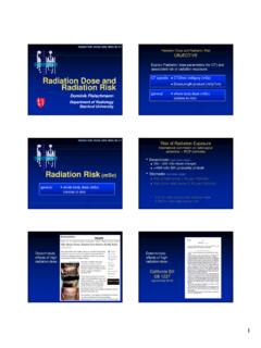
3 Chapter 7. TREATMENT outcomes. The following volumes have been defined as principal volumes related to 3-D TREATMENT PLANNING : gross tumour volume (GTV), CLINICAL target volume (CTV), internal target volume (ITV) and PLANNING target volume (PTV). Figure shows how the different volumes are related to each other. Gross tumour volume The Gross Tumour Volume (GTV) is the gross palpable or visible/. demonstrable extent and location of malignant growth (ICRU Report No. 50). The GTV is usually based on information obtained from a combination of imaging modalities ( computed tomography (CT), magnetic resonance imaging (MRI), ultrasound, etc.), diagnostic modalities (pathology and histological reports, etc.) and CLINICAL examination.
4 CLINICAL target volume The CLINICAL target volume (CTV) is the tissue volume that contains a demonstrable GTV and/or sub- CLINICAL microscopic malignant disease, which has to be eliminated. This volume thus has to be treated adequately in order to achieve the aim of therapy, cure or palliation (ICRU Report No. 50). Organ at risk FIG. Graphical representation of the volumes of interest, as defined in ICRU Reports No. 50 and 62. 220. TREATMENT PLANNING IN EXTERNAL PHOTON BEAM RADIOTHERAPY. The CTV often includes the area directly surrounding the GTV, which may contain microscopic disease and other areas considered to be at risk and requiring TREATMENT ( positive lymph nodes). The CTV is an anatomical . CLINICAL volume and is usually determined by the radiation oncologist, often after other relevant specialists such as pathologists or radiologists have been consulted.
5 The CTV is usually stated as a fixed or variable margin around the GTV ( CTV = GTV + 1 cm margin), but in some cases it is the same as the GTV ( prostate boost to the gland only). There can be several non-contiguous CTVs, which may require different total doses to achieve TREATMENT goals. Internal target volume The ITV consists of the CTV plus an internal margin. The internal margin is designed to take into account the variations in the size and position of the CTV relative to the patient's reference frame (usually defined by the bony anatomy); that is, variations due to organ motions such as breathing and bladder or rectal contents (ICRU Report No. 62). PLANNING target volume The PLANNING target volume (PTV) is a geometrical concept, and it is defined to select appropriate beam arrangements, taking into consideration the net effect of all possible geometrical variations, in order to ensure that the prescribed dose is actually absorbed in the CTV (ICRU Report No.)
6 50). The PTV includes the internal target margin (ICRU Report No. 62) and an additional margin for set-up uncertainties, machine tolerances and intra- TREATMENT variations. The PTV is linked to the reference frame of the TREATMENT machine and is often described as the CTV plus a fixed or variable margin ( PTV = CTV + 1 cm). Usually a single PTV is used to encompass one or several CTVs to be targeted by a group of fields. The PTV depends on the precision of such tools as immobilization devices and lasers, but does not include a margin for the dosimetric characteristics of the radiation beam ( penumbral areas and buildup region), as these will require an additional margin during TREATMENT PLANNING and shielding design.
7 221. Chapter 7. Organ at risk The organ at risk is an organ whose sensitivity to radiation is such that the dose received from a TREATMENT plan may be significant compared with its tolerance, possibly requiring a change in the beam arrangement or a change in the dose. Specific attention should be paid to organs that, although not immediately adjacent to the CTV, have a very low tolerance dose ( the eye lens during nasopharyngeal or brain tumour treatments). Organs with a radiation tolerance that depends on the fractionation scheme should be outlined completely to prevent biasing during TREATMENT plan evaluation. DOSE SPECIFICATION. A clearly defined prescription or reporting point along with detailed information regarding total dose, fractional dose and total elapsed TREATMENT days allows for proper comparison of outcome results.
8 Several dosimetric end points have been defined in ICRU Reports No. 23 and 50 for this purpose: Minimum target dose from a distribution or a dose volume histogram (DVH). Maximum target dose from a distribution or a DVH. Mean target dose: the mean dose of all calculated target points (difficult to obtain without computerized PLANNING ). The ICRU reference point dose is located at a point chosen to represent the delivered dose using the following criteria: The point should be located in a region where the dose can be calculated accurately ( no buildup or steep gradients). The point should be in the central part of the PTV. The isocentre (or beam intersection point) is recommended as the ICRU reference point. Specific recommendations are made with regard to the position of the ICRU reference point for particular beam combinations: For a single beam: the point on the central axis at the centre of the target volume.
9 For parallel opposed equally weighted beams: the point on the central axis midway between the beam entrance points. For parallel opposed unequally weighted beams: the point on the central axis at the centre of the target volume. 222. TREATMENT PLANNING IN EXTERNAL PHOTON BEAM RADIOTHERAPY. For other combinations of intersecting beams: the point at the inter- section of the central axes (insofar as there is no dose gradient at this point). PATIENT DATA ACQUISITION AND SIMULATION. Need for patient data Patient data acquisition is an important part of the simulation process, since reliable data are required for TREATMENT PLANNING purposes and allow for a TREATMENT plan to be properly carried out. The type of gathered data varies greatly, depending on the type of TREATMENT plan to be generated ( manual calculation of parallel opposed beams versus a complex 3-D TREATMENT plan with image fusion).
10 General considerations include: Patient dimensions are almost always required for TREATMENT time or monitor unit (MU) calculations, whether read with a calliper, from CT. slices or by other means;. The type of dose evaluation dictates the amount of patient data required ( DVHs require more patient information than a point dose calculation of organ dose);. Landmarks such as bony or fiducial marks are required to match positions in the TREATMENT plan with positions on the patient. Nature of patient data The patient information required for TREATMENT PLANNING varies from rudimentary to very complex, ranging from distances read on the skin, through manual determination of contours, to acquisition of CT information over a large volume, or even image fusion using various imaging modalities.