Transcription of CHARACTERISTICS OF PROKARYOTIC AND EUKARYOTIC CELLS
1 CHARACTERISTICS OF PROKARYOTIC AND EUKARYOTIC CELLS CHAPTER 4 Detailed studies of CELLS have revealed that prokaryotes differ enough to be split into two large groups called domains. A relatively new concept in classification, domain is the highest taxonomic category, higher even than kingdom. Three domains exist: Archaea (archaeobacteria) Bacteria (eubacteria) Eukarya Archaea and Bacteria are both prokaryotes. The comparison in the next few slides is between bacteria and EUKARYOTIC CELLS . We will deal with archaea in a following slide. Basic Cell Types: PROKARYOTIC are CELLS that lack a nucleus (nuclear membrane). Prokarotic CELLS are single CELLS but are subdivided into Bacteria and Arachaea as mention in the previous slide.
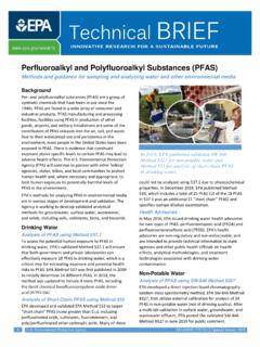
2 EUKARYOTIC CELLS contain a nucleus (nuclear membrane). EUKARYOTIC CELLS include: plants, animals, fungi and protists ( a very heterogeneous group that are neither animals, plants or fungi and are often single cell and small , protozoa). Prokaryotes (Bacteria) and Eukaryotes have many similarities and many differences: Cellular CHARACTERISTICS Cellular CHARACTERISTICS Size: Most prokaryotes range from to m in diameter. A human red blood cell is about m. Fig. The most common bacterial shapes Shape Bacteria which show a wide variety of shapes within a single species are said to be Pleomorphic. Those of you taking the laboratory will see the various shapes of bacteria first-hand. Fig. Arrangements of some of The more common cocci forms of bacteria An overview of structure Fig.
3 A typical PROKARYOTIC cell 1. Cell membrane-typically surrounded by a cell wall 2. Internal cytoplasm with ribosomes, a nuclear region and granules 3. Lots of external structure, , capsules, flagella and pili Capsule stain Endospore stain Acid fast stain Gram stain For those of you not in the laboratory these videos will show you how the stains are done and how stained CELLS appear. Pili and bacterial attachment View on your own The Cell Wall The semi-rigid cell wall lies outside the cell membrane in nearly all bacteria (Mycoplasma being an exception). A. maintains cell shape B. acts as a corset in preventing CELLS from bursting in hypotonic solution (you can store a culture of E. coli in distilled water for weeks to months) C.
4 It is quite porous and has little effect on the inflow and outflow of materials Peptidoglycan- the structure that gives shape and strength to bacteria. Many bacteria can be stored in distilled water because that have a peptidoglycan "corset". Gram positive and Gram negative organisms have somewhat different peptidoglycan layers as we will see on the next slide. The peptidoglycan layer permits bacteria, , E. coli to be stored in hypotonic solutions, , distilled water, for weeks. Fig. A-two dimensional view of Gram negative wall, Gram negative peptidoglycan (E. coli) which is basically a two-dimensional structure because it lacks the pentaglycine cross-linker of Gm + orgs. Fig. A three-dimensional view of peptidoglycan for Gram positive bacteria Synthesis of peptidoglycan Although we will deal with antibiotics that block cell wall synthesis in another chapter- having seen how the cell wall is synthesized how penicillin block cell wall synthesis would be in order (even though we will see it again in the antibiotic chapter).
5 How penicillin inhibits cell wall synthesis Periplasmic space - A gap between the cell wall and the cytoplasmic membrane most easily observed in Gram negative organisms since they have a double membrane and is not characteristically considered a feature of gram positive organisms although some refer to the space between inner and outer leflets of the plasma membrane of Gram positive organisms as a periplasmic space (I do not). It is a region of high enzymatic activity containing many digestive enzymes and transport proteins. It is also a region that is outside of the normal array of digestive enzymes associated with the cytoplasm (in particular certain proteolytic enzymes). We will see it in fig. Distinguishing Bacteria by Cell Walls- Gram negative and Gram positive Peptidoglycan layer Gm + and Gm - Fig.
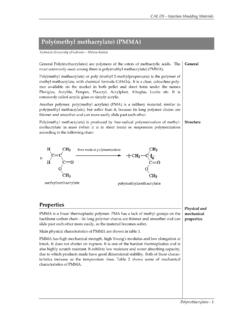
6 Gram positive cell wall * Teichoic acid a polymer of glycerol, phosphate and ribitol occurs in units up to 30 units long extends beyond the cell wall Only found in Gm + organisms, but not all Gram + organism contain teichoic acid. Gram Negative Cell Walls Outer Membrane Periplasmic space Digestive Enzymes Protein pumps LPS components Fig. Gram negative cell wall Acid Fast Cell Wall Fig. fast cell wall Mycobacterium tuberculosis (TB organism is an acid fast organism ) Lipid bilayer peptidoglycan arabinogalactan Mycolic acid Acyl lipids lipoarabinomannan (LAM) The complex structure of the acid fast cell wall makes it a good barrier against many physical agents, such as phagocytic cell digestion, penetration by antibacterial agents.
7 Lipoarabinomannan (LAM) is a glycolipid, and a virulence factor associated with Mycobacterium tuberculosis, the bacteria responsible for tuberculosis. Its primary function is to inactivate macrophages and scavenge oxidative radicals. Mycolic acid is also associated with the virulence of M. tuberculosis. * PROKARYOTIC Plasma Membrane Phospholipids Fluid Mosaic Novel Stem Cell treatment May Hold Promise for Type 1 Diabetes) -- A new type of stem cell treatment for people with type 1 diabetes appears to help re-educate rogue immune system CELLS , which allows CELLS in the pancreas to start producing insulin again. The treatment , which combines a patient's immune system CELLS with stem CELLS from a donor's cord blood, even worked in people with long-standing diabetes who were believed to have no insulin-producing ability.
8 Although the treatment didn't wean anyone off insulin completely, average blood sugar levels dropped significantly, which would reduce the risk of long-term complications. "Our study brings a new hope for people with type 1 diabetes. If we can control the autoimmunity, we may reverse the diabetes. We showed that the islets [ CELLS ] can start to work again," said Dr. Yong Zhao, an assistant professor in the section of endocrinology, diabetes and metabolism at the University of Illinois at Chicago. The study participants, who were 15 to 41 years old, had had type 1 diabetes for an average of nine years. Six had some residual beta cell function and six did not. Both groups were given stem cell educator therapy. The other three people served as the control group.
9 The researchers measured C-peptide, a protein fragment that's a byproduct of insulin production, and found that the educator therapy group had improved levels of C-peptide at 12 weeks. These levels continued to improve until 24 weeks, and remained stable through the follow-up at 40 weeks. There were no changes in C-peptide in the control group. The average daily dose of insulin dropped almost 39 percent after 12 weeks for the group with some beta cell function and 25 percent in those with no beta cell function, suggesting that the group with no beta cell function now produced insulin. That means if you stop the autoimmune reaction, you may see beta cell regeneration, or there might be other precursor CELLS in the pancreas. If these data are confirmed, this is a very provocative and remarkable finding," Inverardi said.
10 The average hemoglobin A1C level dropped percent for those with residual beta cell function and percent for those without beta cell function. A1C levels measure average blood sugar levels over two to three months, and people with type 1 diabetes are advised to maintain A1C levels below 7 percent. A drop of 1 percent in A1C levels can reduce the risk of complications. This was an initial clinical trial designed to test for safety. Zhao said that in future trials he hopes that with additional treatments people might get off insulin altogether. Fig. The fluid-mosaic model of the cell membrane- Several reasons why this generic structure looks more like a EUKARYOTIC plasma membane than a PROKARYOTIC one The Cytoplasm-semi-fluid substance inside the cell membrane.