Transcription of Cholangiocarcinoma: Introduction
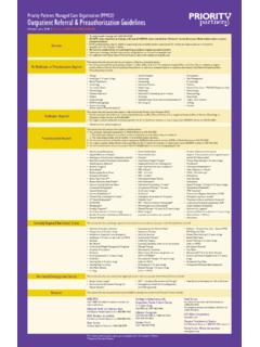
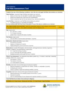
1 Figure 1. Location of the biliary tree in the : Introduction The terms cholangiocarcinoma and bile duct cancer are often used interchangeably. Primary biliary tract malignanciesaffect one in every 100,000 people per year in the United States. More than 95% of these malignancies arecholangiocarcinomas (epithelial adenocarcinomas ) frequently found in the extrahepatic biliary tree. This form of canceris slightly more prevalent in males than females ( ) and usually affects patients in the fifth to seventh decade of life. What is Cholangiocarcinoma?Cholangiocarcinoma is a primary malignant tumor originating from cells that resemble biliary epithelium. The gross appearance is that of one or more firm, whitemasses (Figure 2). Figure 2. A, Extrahepatic tumor; B, intrahepatic tumor resulting in biliary duct dilation. The tumor(s) is usually small and may arise anywhere along the biliary tree, from the small intrahepatic bile ducts to the common bile duct. Microscopically,cholangiocarcinoma may resemble adenocarcinoma.
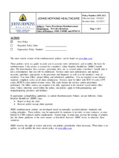
2 These bile ductule tumors may be well differentiated, while others are poorly differentiated (Figure 3). Figure 3. A, Cholangiocarcinoma within a cirrhotic liver; B, cross-section of tumor; C, histologicalimaging showing tumor cells surrounding normal hepatocytes. Cholangiocarcinomas are usually slow-growing tumors that spread locally via the lymphatic system. Treatment and long-term prognosis are dependent upon thelocation of the mass. Lesions located in the distal or middle portion of the extrahepatic bile duct (20% and 35%, respectively) have a better prognosis than tumors inthe proximal third, which include about 45% of bile duct cancers (including Klatskin s tumors hilar variants).Large solitary tumors are characteristic of peripheral cholangiocarcinoma; however, a multinodular type may occur. These tumors have a fibrous stroma, are firm andgrayish white in color, and are not well vascularized. Hilar cholangiocarcinoma are usually firm, intramural, annular tumors that encircle the bile duct, or may be bulkyhard masses that are on the duct or hilar region and extend into the liver.
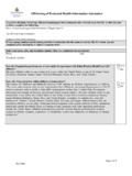
3 They may also appear as a spongy friable mass in the lumen of the bile duct. There may bemetastatic nodules throughout the liver with dilation of bile ducts peripheral to the mass. SymptomsThe clinical presentation of cholangiocarcinoma depends on the anatomic location of the tumor(s). Patients with hilar cholangiocarcinoma, (tumor located in the areaof confluence of right and left hepatic ducts) most commonly present with jaundice, pruritis, abdominal pain, fever, weight loss and/or progressive weakness (Figure4). Patients with peripheral cholangiocarcinoma (tumor originating from small intrahepatic ducts) may present only with vague abdominal pain, unexplained weightloss, weakness and worsening fatigue. Jaundice and pruritus may not be apparent until very late in the disease course, when there is occlusion of segmental bileducts. Patients with distal cholangiocarcinoma (tumors involving extrahepatic bile ducts) usually have early onset of jaundice and pruritus without abdominal physical examination, these patients usually have a palpable distended gallbladder (Courvoisier s sign).
4 Figure 4. Common symptoms of may occur in the setting of primary sclerosing cholangitis and may be difficult to diagnose. Clues that may suggest an underlying carcinomainclude a stricture that is refractory to therapy, or sudden deterioration in biochemical tests of liver function. Copyright 2001-2013 | All Rights North Wolfe Street, Baltimore, Maryland 21287 Cholangiocarcinoma: anatomy anatomy AnatomyThe liver arises from the ventral mesogastrium, and only the upper posterior surface is outside of that structure. The ligamentum teres and falciform ligament connectthe liver to the anterior body wall. The lesser omentum connects it to the stomach and the coronary and triangular ligaments to the diaphragm. The liver is smooth andfeatureless on the diaphragmatic surface and presents with a series of indentations on the visceral surface where it meets the right kidney, adrenal gland, inferiorvena cava, hepatoduodenal ligament and stomach .The liver can be considered in terms of blood supply, hepatocytes, Kupffer cells and biliary passages.
5 The liver receives its blood supply from the portal vein andhepatic artery, the former providing about 75% of the total 1500 mL/min flow. Small branches from each vessel (the terminal portal venule and terminal hepaticarteriole) enter each acinus at the portal triad (Figure 6). Pooled blood then flows through sinusoids between plates and hepatocytes exchanging nutrients. Thehepatic vein carries all efferent blood into the inferior vena cava, and a supply of lymphatic vessels drains the 6. A, Histological illustration showing a magnified view of the portal tract; B, liver lobuleLiver cells, or hepatocytes, comprise the bulk of the organ, which carry out complex metabolic processes. Hepatocytes are responsible for the liver s central role inmetabolism. The functions of these cells include the formation and excretion of bile, regulation of carbohydrate homeostasis, lipid synthesis and secretion of plasmalipoproteins, control of cholesterol metabolism, formation of urea, serum albumin, clotting factors, enzymes and numerous proteins.
6 The liver also aids in themetabolism and detoxification of drugs and other foreign cells line the hepatic sinusoid and are part of the reticuloendothelial system filtering out minute foreign particles, bacteria and gut-derived toxins. They alsoplay a role in immune processes involving the passages begin as tiny bile canaliculi formed by hepatocytes. These microvilli-lined structures progress into ductules, interlobular bile ducts and larger hepaticducts. Outside the porta hepatis, the main hepatic duct joins the cystic duct from the gallbladder to form the common bile duct, which drains into the duodenum. Copyright 2001-2013 | All Rights North Wolfe Street, Baltimore, Maryland 21287 Cholangiocarcinoma: Causes Primary Sclerosing CholangitisThere is a high incidence of cholangiocarcinoma in patients (Figure 7) with ulcerative colitis (1 in 256) and primary sclerosing cholangitis (4 20%). The cumulative riskfor cholangiocarcinoma is at 10 years after 7.
7 A, Klatskin s tumor (tumor located in the hepatic duct bifurcation) in a patient with primarysclerosing cholangitis; B, corresponding cholangiogram (ERCP image). Liver Flukes Cholangiocarcinoma is more common in areas endemic to liver fluke infection (Hong Kong, Thailand). Liver flukes, such as Clonorchis sinensis or Opisthorchisviverrini, usually enter human s gastrointestinal tract after ingestion of raw fish. Parasites travel via the duodenum into the host s intrahepatic or extrahepatic biliaryducts. Liver flukes cause bile stasis, inflammation, periductal fibrosis and hyperplasia, with the subsequent development of cholangiocarcinoma (Figure 8). Figure 8. A, Liver flukes; B, micrograph of liver fluke eggs in the liver (reused with permission: Sun etal., Ann. Clin. Lab. Sci., 1984). GallstonesGallstones vary in size, shape and number, and may be found throughout the biliary tract. The link between cholangiocarcinoma and gallstones is gallstones may cause chronic obstruction to bile flow, promote micro injury of the bile ducts, and are associated with a 2 10% risk of the development ofcholangiocarcinoma (Figure 9).
8 Congenital cystic dilation of intrahepatic biliary ducts (Caroli s disease), and choledochus cysts have also been closely associated withdevelopment of cholangiocarcinoma. Figure 9. Intrahepatic biliary gallstones resulting in ductal dilation. Thorotrast The radiocontrast agent, Thorotrast, was in use from the late 1920s through the 1950s. There are many reports of development of cholangiocarcinoma 30 35 yearsafter exposure to this contrast material. Copyright 2001-2013 | All Rights North Wolfe Street, Baltimore, Maryland 21287 Cholangiocarcinoma: Diagnosis Laboratory testsBiochemical tests of liver function may reveal a cholestatic picture with elevated total bilirubin and alkaline phosphatase. This pattern is non-specific forcholangiocarcinoma and may be found with any cause of obstruction to bile flow. The levels of blood bilirubin and alkaline phosphatase usually correlate with degreeand duration of obstruction of the biliary ducts. Fluctuation in the serum bilirubin level may reflect incomplete obstruction and involvement of one hepatic and CA19-9 Carcinoembriogenic antigen (CEA) and CA 19-9 are blood tests for non-specific markers of underlying gastrointestinal malignancies.
9 These tests are positive in morethan 40% of patients with cholangiocarcinoma, but usually only in late stages of the (AFP)Alpha-fetoprotein is another blood test commonly used to identify markers of possible hepatobiliary malignancy. This test is usually elevated in patients withcholangiocarcinoma, but not to the degree of elevations in patients with hepatocellular carcinoma. Radiological DiagnosisUltrasoundTransabdominal ultrasound is a totally painless, non-invasive procedure. The test does not require special preparation, although it is technically easier in patients withat least six hours of fasting. Transabdominal ultrasound is usually recommended as the first imaging modality for the investigation of patients with suspectedcholangiocarcinoma. In hilar cholangiocarcinoma, ultrasound demonstrates bilateral dilation of intrahepatic ducts, and right and left hepatic ducts. In rare cases, thetumor itself can be visualized as either a hypoechoic (decreased echodensity) or hyperechoic (increased echodensity) rounded mass located just distal to dilatedbiliary ducts.
10 Peripheral cholangiocarcinoma may be suspected if abdominal ultrasound demonstrates local dilation of intrahepatic ducts or isolated dilation of thebiliary tree inside one lobe of the liver. In both peripheral and hilar cholangiocarcinoma, biliary ducts distal to the obstruction (common hepatic duct and common bileduct) are not dilated. In patients with hilar cholangiocarcinoma and complete obstruction of both right and left hepatic ducts, extrahepatic bile ducts and thegallbladder appear empty (collapsed) because there is no bile flow out of the liver. In patients with distal cholangiocarcinoma, ultrasound demonstrates dilated intra-and extrahepatic ducts along with significant dilation of the gallbladder. Peripherally located tumors cause segmental or lobular obstruction of the biliary tree. Bile flowfrom the rest of the liver is preserved. Extrahepatic bile ducts and the gallbladder appear normal (filled with bile) in patients with peripheral ultrasound can also detect the presence of liver metastases as single or multiple rounded lesions of different Tomography (CT)Computed tomography may detect lesions of low-density mass associated with dilated biliary ducts (Figures 10 and 11).