Transcription of Chondral Lesions of the Hip - Arthrosurface
1 Chondral Lesions of the HipMicrofracture and ChondroplastyYi-Meng Yen, MD, PhD and Mininder S. Kocher, MD, MPHA bstract:Hip arthroscopy has become increasingly popular overthe past several years as the techniques have evolved to be ableto address both the peripheral and central compartments ofthe hip. The main indications for hip arthroscopy 10 years agowere diagnostic and debridement procedures such as removalof loose bodies, labral resection, synovectomy, and cartilagedebridement. Advances in this field have now expanded to includereconstruction and repair of the labrum, recontouring of theacetabulum and head-neck junction, cartilage salvage, and repairand releases of the tendons around the hip joint. We detail in thisarticle Chondral injuries that occur in the hip joint and arthroscopicprocedures to address these issues.
2 We routinely perform chon-droplasty in cases where there is a partial thickness tear of articularcartilage. Full thickness defects are addressed with microfracturewhich follows closely the guidelines established for the knee. As ourunderstanding of Chondral injuries and their causes grows, futureefforts will focus on :articular cartilage, injury, arthroscopy(Sports Med Arthrosc Rev2010;18:83 89) Chondral injuries of the hip can be an elusive source ofjoint pain. Although radiographic diagnosis is improv-ing, even gadolinium-enhanced arthrography is limited inits ability to reliably show the presence and extent of may be explained by the static nature ofimaging the joint and lack of distraction. More recently,however, delayed gadolinium-enhanced magnetic resonanceimaging (MRI) of cartilage has been in use to delineatechondral damage within the hip with good injuries of the hip can be observed in avariety of hip disorders and can result from either anatraumatic or traumatic etiology.
3 These hip disorders includeloose bodies from Perthes or other chondrodysplasias, hipinstability or dislocation, osteonecrosis of the femoral head,slipped capital femoral epiphysis, hip dysplasia, femoroace-tabular impingement (FAI), and degenerative joint disease,amongst others. Chondral injuries can be acute, chronic, ordegenerative and may involve a full or partial arthroscopy has become increasingly popular overthe past several years as indications have shifted from adiagnostic tool to a therapeutic one. It is known thatarticular cartilage defects have limited healing capacityregardless of the acute, chronic, or degenerative nature ofthe such, the role of hip arthroscopy is shiftingin some regards to the prevention of articular cartilageinjuries as observed in FAI. However, the treatment ofexisting cartilage injuries of the hip has mainly beenadapted from studies on the knee.
4 These techniques includechondroplasty, abrasion arthroplasty, osteochondral dril-ling, osteoarticular autograft or allograft, hemicap resurfa-cing,4autologous chondrocyte implantation (ACI), 7 With the exception of osteoarticulargrafts, these techniques produce a fibrocartilage and nota true articular cartilage surface. The use of ACI andosteoarticular grafting has only been described using ,9 The microfracture technique of the knee asdescribed by Steadman et al10 12has becoming increasinglypopular as a preferred treatment method for chondraldefects with good results. Microfracture is a marrow-stimulating procedure that brings undifferentiated stemcells from a subchondral perforation into the chondraldefect. A clot formed in the microfractured area providesan environment for both pluripotent marrow cells andmesenchymal stem cells to differentiate into stable fibro-cartilaginous INJURIESL oose BodiesLoose bodies are free-floating fragments usually ofcartilage, and arthroscopic removal of these loose bodieshas been one of the classic indications for hip loose bodies can originate from either traumaticinjury, degenerative changes, or other disease processessuch as Perthes, spondyloepiphyseal dysplasia, or osteo-necrosis.
5 Loose bodies are commonly found in the posterioraspect of the central compartment and/or in the peripheralcompartment, especially in the posterior and medialfemoral neck of the femoral head can give rise toloose bodies and articular cartilage defects. Hip arthro-scopy can be useful for the removal of the associated loosebodies, chondroplasty, or microfracture of the articularcartilage and drilling on the avascular zone under and Jones14treated 4 patients withavascular necrosis of the femoral head and reported theworst results in 3 patients with the most advanced DissecansOsteochondritis dissecans of the hip joint has beenreported in the femoral head and the 18 Hiparthroscopy can be used to remove the osteochondralCopyrightr2010 by Lippincott Williams & WilkinsFrom the Children s Hospital, Boston, The Adolescent and YoungAdult Hip Unit, Department of Orthopaedic Surgery, Division ofSports Medicine, Boston, : Mininder S.
6 Kocher, MD, MPH, Children s Hospital,Boston, Division of Sports Medicine, The Adolescent and YoungAdult Hip Unit, Department of Orthopaedic Surgery, 300 Long-wood Avenue, Boston, MA 02115 (e-mail: Med Arthrosc Rev Volume 18, Number 2, June |83fragment and debride and/or drill the resultant lesion . Thisdisease is rare and the successful treatment with hiparthroscopy was only reported in few case ,17 FAITwo different types of FAI are recognized, cam andpincer, which lead to characteristic cartilage injury impingement results from pathologic contactbetween an abnormally shaped femoral head and neck witha normal acetabulum. As the hip flexes, particularly ininternal rotation, this abnormal region of the proximalfemur contacts the acetabulum. The resultant shear forcescauses acetabular cartilage damage, including delamination(termed the carpet or wave phenomenom20) and frankgrade IV chondromalacia.)
7 In severe cases of cam impinge-ment, the labrum will become torn. The location of cartilageinjury is the anterosuperior region of the impingement is a result of abnormal contactbetween the acetabular rim which is either anteriorlylocalized (acetabular retroversion) or generally deep (coxaprofunda or acetabular protrusion) with a typically normalfemoral head-neck junction. Repeated compressive forcesin this area lead to failure of the acetabular labrum. Thebase of the labrum tends to develop intralabral ganglions orossifications. The region of Chondral injury is more shallowthan that of strict cam impingement, but usually encom-passes a more global rim around the superior ,22 Repeated impingement of the anterior femoralhead on the acetabulum results in a moment arm on theposteroinferior acetabulum and the so-called contre-coup-cartilage injury of the posteroinferior AND PHYSICAL EXAMINATIONThe history and physical examination of the hip isthe most useful and effective way to diagnose and treat hipdisorders.
8 The clinician must be able to determine thefrequency, severity, location, inciting factors, and radiationof symptoms. Mechanical symptoms such as clicking andlocking can be seen with labral tears, audible snapping maybe associated with coxa saltans interna or externa. Factorsthat exacerbate or relieve the symptoms and whether thesymptoms are chronic or acute can aid in the hip pain usually presents as groin pain andmay radiate to the anterior thigh, although patientsdescribe pain on the lateral thigh or even buttocks,oftentimes placing their hands cupped over their greatertrochanter, the C-sign. 23,24 Pain from lateral thigh andbuttocks must also be investigated for a ,26 Unless the Chondral Lesions are in the form ofloose bodies, pure Chondral injuries may not present withmuch pain or symptoms as cartilage does not havenociceptive receptors.
9 However, synovial irritation fromchondral damage would present as a painful physical examination should be thorough. Thepatient s gait should be noted and any leg-length discre-pancies assessed. Examination of the lumbar spine includ-ing motor function, sensation, range of motion, reflexes,and straight-leg raises must be performed to rule outlumbar spine pathology as the cause of symptoms. The hipexamination begins with palpation of bony prominencesabout the hip and assessment of range of motion. Side-to-side differences should be noted as they may indicateareas of pathology. Clicking, catching, or other mechanicalsymptoms during the examination should be noted. Althoughchondral injuries may be associated with mechanicalsymptoms, there is no specific examination maneuver toassess for them. The impingement test should be performedin the supine radiographs are the most useful imaging tool forthe initial evaluation of hip complaints.
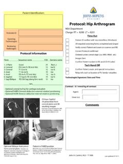
10 Radiographs canshow degenerative changes, bony Lesions , dysplasia, thepresence of osteochondral loose bodies, and evaluation should include an anteroposterior(AP) pelvis, a lateral radiograph, and false profile there has been shown to be no difference inweight-bearing surface of an AP pelvis taken supine versusstanding,27a standing AP pelvis can accentuate evidenceof mild dysplasia. Furthermore, morphologic change of theacetabulum such as retroversion, coxa profunda, andprotrusion acetabuli can be identified. The lateral radiographis useful for examination of the anterolateral head-neckjunction whereas the false profile views give informationabout anterior coverage of the acetabulum. In addition,computed tomography can be performed to aid in an MRI allows improved visualization ofthe soft tissues, early degenerative changes, and osteone-crosis,28it has been shown earlier that plain MRI does notaccurately identify labral or Chondral defects, althoughthere are newer imaging protocols that are more ,30 MRI arthrogram can allow for improved visua-lization of intra-articular gadolinium isinjected into the hip, which distends the capsule and allowsbetter visualization of the articular cartilage and Lesions can be better visualized when the cartilagedefect is outlined by gadolinium.