Transcription of Clinical Biomechanics of the Spine White AA, Panjabi MM ...
1 Clinical Biomechanics of the Spine White AA, Panjabi MM. 1990, ISBN 0-397-50720-8 Second Edition Auszug: seite 18 und 19. Mechanisms of Disc Prolapse By using fresh cadaveric lumbar Spine segments, many attempts have been made to produce a disc prolapse that is similar to that seen clinically. Compressive loading of the disc, at slow speed or at high speed, is known to result in fractures of the endplates and not in failure of the disc. 93,203,254. This is true irrespective of the status of disc degeneration. 67 Torsional loading of the disc beyond its physiologic limits does result in circumferential tears in the annulus but does not result in disc prolapse. 67 Simple flexion of the specimen generally results in tearing of the posterior ligaments 215.
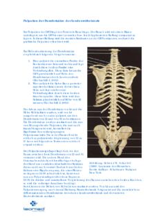
2 Or fractures of the laminae. Therefore, the experiments described below are of significant Clinical importance. For the first time, disc prolapse was produced in a laboratory setting with the use of fresh cadaveric specimens. The disc prolapse was attempted by sudden load applications as well as by gradual loading. Sudden Disc Prolaps The two-vertebrae specimen, laminectomized for observation of disc prolapse, was positioned such that the upper vertebra was laterally bent and hyperflexed so that the posterolateral aspect of the disc annulus was under tension. 3 This was on the side opposite the side of the lateral bend. In this set posture the specimen was suddenly loaded by a compressive force. The result was disc prolapse, similar to that seen clinically, in 26 of the 61 specimens tested.
3 A close scrutiny of the specimens showed that there was a certain pattern to the specimens that prolapsed and the ones that did not. The prolapse-prone specimens most likely came form the lower lumbar levels (L4-L5 or L5-S1), the 40-49-year age group, and disc degeneration with a grade of 2 (on a scale of 1-4) (Fig. 1-12). These sets of attributes seem to correlate well with the Clinical picture of disc prolapse. In addition, these attributes also coincide with the instability stage of the degeneration hypothesis of Kirkaldy-Willis. 118 In his hypothesis, the first stage of degeneration may result in some spinal dysfunction but no instability. In the third stage, the Spine is restabilized, probably because of ligament calcification and osteophytes.
4 However, in the second stage, which occurs between the ages of 40 and 50 years, the disc degeneration has progressed to the point where the nucleus is still mobile. This is the instability stage. At this stage there is increased risk of disc prolapse at the L4-L5 or L5-S1 levels because of traumatic overload of the Spine . Figure 1-12 A mechanism of sudden disc prolapse. Using fresh cadaveric lumbar Spine specimens, with posterior elements removed for observation of the disc, experimental disc prolapse was produced in 43% of the experimental trials. The method consisted of placing the specimen in a fully flexed and somewhat laterally bent posture (thus producing tension in the annular fibers) and applying a sudden compression load. The prolapse was produced on the side opposite the side of the lateral bend.
5 Most susceptible discs were those at L5-S1, 40-50 years old, and with a degeneration grade of 2 (on a scale of 1 to 4). (Data from Adams, M. A., and Hutton, W. C.: Prolapsed intervertebral disc. A hyperflexion injury. Spine , 7: 184, 1982). Gradual Disc Prolaps Since the majority of the low back pain patients with disc prolapse seen clinically do not report a precipitating traumatic event, an attempt was made to produce disc prolapse in the laboratory using slowly varying loads. 6 Fresh cadaveric lumbar FSUs were positioned so that the application of axial compression resulted in simultaneous compression, flexion, and some lateral bending. Cyclically varying compression, between 1500 and 6000 N, was applied at a rate of 40 loadings per minute.
6 Of the 49 specimens tested, only 6 had gradual prolapses, 35 had end-plate fractures or vertebral collapse, and 8 did not fail at all. One may conclude that the gradual disc prolapse is most likely not caused by the hyperflexion loading used in this experiment. It may be the result of a combination of factors, such as weakened posterior disc annulus, relatively degenerated annulus with fissures, and another kind of loading (e. g., bending and twisting). Auszug: seite 106. The Lumbar Spine Range of Motion The representative rotations in flexion/extension, lateral bending, and axial rotation are shown in Table 2-4 and Figure 2-23. In flexion/extension there is usually a cephalocaudal increase in the range of motion in the lumbar Spine . The lumbosacral joint offers more sagittal plane motion than do the other lumbar joints.
7 For lateral bending, each level is about the same, except for the lumbosacral joint, which shows a relatively small amount of motion. The situation is about the same for axial rotation. 66 It is not unreasonable to speculate that the high incidence of clinically evident disc disease at L4-L5 and L5-S1 may be related to mechanics. These two areas bear the highest loads and tend to undergo the most motion in the sagittal plane. An important component of lumbar Spine kinematics is that of sagittal plane translation. This is because measurement of this parameter is frequently used to determine whether or not there is instability. There is considerable variation in measuring techniques. The work of Pearcy 95 is based on sound methodology and suggests that 2 mm of anterior sagittal plane translation is normal for the lumbar Spine .
8 The in vitro work of Posner and colleagues, 97 who used preloads to simulate physiologic conditions, suggested mm of anterior displacement as the upper limits of normal. Thus, after careful consideration of a number of factors, we suggest mm for evaluation of Clinical instability (see p. 354). Auszug: seite 354. Measurements can be made directly from resting or flexion/extension radiographs. In the acute traumatic setting, resting radiographs are usually performed. Sagittal plane displacement greater than mm or 15% of the anteroposterior diameter of the vertebral body on a static (resting) lateral radiograph should be considered potentially unstable. These values were obtained from the aforementioned biomechanical experiment. 125 Relative sagittal plane angulation greater than 22 is abnormal and potentially unstable at any level in the lumbar Spine .
9 Note that 22 of relative angulation means 22 greater than the amount of angulation at the FSU above or below the FSU in question (Fig. 5-62). These norms were obtained from a review of the literature of the normal resting sagittal posture of the lumbar Spine . 14a,145a This value was tested on the data of 102 normal subjects. This standard of comparison takes into account the normal angulation between FSUs. Figure 5-61 Measurement to determine vertebral translation or displacement in the lumbar Spine . A method for measuring sagittal plane translation or displacement. If the translation or displacement is as much as mm or 15 of the sagittal diameter of the adjacent vertebra, it is considered to be abnormal. These measurements are to be used in conjunction with the checklist in Table 5-10.
10 After evaluation with resting radiographs in the acute traumatic setting or in the nonacute setting of evaluating for lumbar Spine Clinical instability, additional information may be gained by obtaining flexion/extension radiographs. Sagittal plane translation greater than mm or 15% of the anteroposterior diameter of the vertebral body on dynamic (flexion/extension) radiographs should be considered potentially unstable. These values were obtained from the aforementioned experimental study and several other kinematic studies. 44a,66a,114a,125,173a Sagittal plane rotation on dynamic radiographs greater than 15 at L1-L2, L2-L3, and L3-L4, greater than 20 at L4-L5, or greater than 25 at L5-S1 is abnormal and potentially unstable (Fig. 5-63). These values were based on a review of the literature of in vitro and in vivo lumbar Spine ranges of motion.